The 14 Hallmarks of Aging: How NAD+ Plays a Role in Every Hallmark
Key Takeaways
- Aging is a system of biological processes, not isolated events: 14 biological and psychological/social hallmarks interact to drive the aging process.
- NAD+ is a central hub: A decline in NAD+ affects multiple hallmarks, from mitochondrial health to DNA repair, stem cell function, and inflammation.
- Protective processes can backfire: Responses like autophagy and cellular senescence are initially beneficial but can become harmful over time.
- New hallmarks expand our understanding: Extracellular matrix alterations and socio-psychological isolation highlight the structural and social dimensions of aging.
- Interventions should be multi-pronged: Supporting NAD+ metabolism, alongside lifestyle, social, and mental health strategies, may create ripple effects across aging pathways.
Aging is not simply the passage of time or the number of candles on your birthday cake each year; rather, it is a reflection of how well—or poorly—your body is functioning on a cellular and molecular level.
This reflection is seen in our biological age, which is often different from our chronological age. While chronological age measures time in a standard 365-day year, biological age reflects the cumulative impact of molecular damage in your body—essentially, how fast your cells, organs, and tissues are aging. And while these two tend to line up during our younger years, as we grow older, they can drift further and further apart.
With a biological age that greatly exceeds your chronological age, you’re more likely to experience accelerated aging and an earlier onset of age-related disorders. But when chronological and biological age are in sync, this is a reflection of dynamic equilibrium, or the body’s ability to maintain a stable internal environment despite ongoing stressors. According to the World Health Organization (WHO), aging is an accumulation of molecular and cellular damage over time—essentially, a disruption to dynamic equilibrium that accelerates damage and physiological decline.
Understanding why aging occurs in the first place is the first step in slowing it down. A major advancement in this area came with the "hallmarks of aging" framework, first proposed by Carlos López-Otín and his team at the University of Oviedo in Spain.¹ This framework identifies the main biological processes that drive aging, helping us understand how cellular damage builds up over time.
In this article, we’ll explore the currently proposed 14 hallmarks of aging, including the underlying mechanisms behind each, and highlight how cellular metabolism—especially nicotinamide adenine dinucleotide (NAD+)—interacts with these processes.
What Are the 14 Hallmarks of Aging? The Evolving Scientific Framework
First described as a set of nine characteristics in a 2013 paper published in Cell, the hallmarks of aging are fundamental characteristics that define why our bodies age.¹ A decade later, these nine hallmarks were expanded to 12,² and most recently, in 2025, extended to 14,³ with the addition of extracellular matrix (ECM) alterations and socio-psychological isolation.
This evolution reflects a more holistic understanding of aging. While early research focused more on biological mechanisms, it’s now recognized that psychological and social factors also play a vital role in why aging occurs. It’s important to recognize the principle of interdependence, as amplifying or reducing a singular hallmark typically does not act in a vacuum and will affect the others. Aging must be understood as a whole system, not a collection of 14 isolated problems to fix. At the same time, each hallmark corresponds to measurable molecular or cellular changes and represents a potential point for intervention.
The hallmarks can be grouped into three layers: primary, antagonistic, and integrative hallmarks. Primary hallmarks represent root-cause molecular damage, or the body’s “wear and tear.” These include genomic instability, telomere attrition, epigenetic alterations, and loss of proteostasis. Antagonistic hallmarks are initially protective responses that can become harmful over time, including deregulated nutrient sensing, mitochondrial dysfunction, cellular senescence, and disabled macroautophagy. Integrative hallmarks reflect downstream systemic effects, typically the “visible” consequences of aging: stem cell exhaustion, altered intercellular communication, chronic inflammation, dysbiosis, ECM alterations, and socio-psychological isolation.
Understanding these layers and how they interact with one another sets the stage for how cellular metabolism—and specifically, NAD+—interacts with multiple hallmarks simultaneously to potentially slow aspects of aging.
NAD+ and the Hallmarks of Aging: A Central Thread
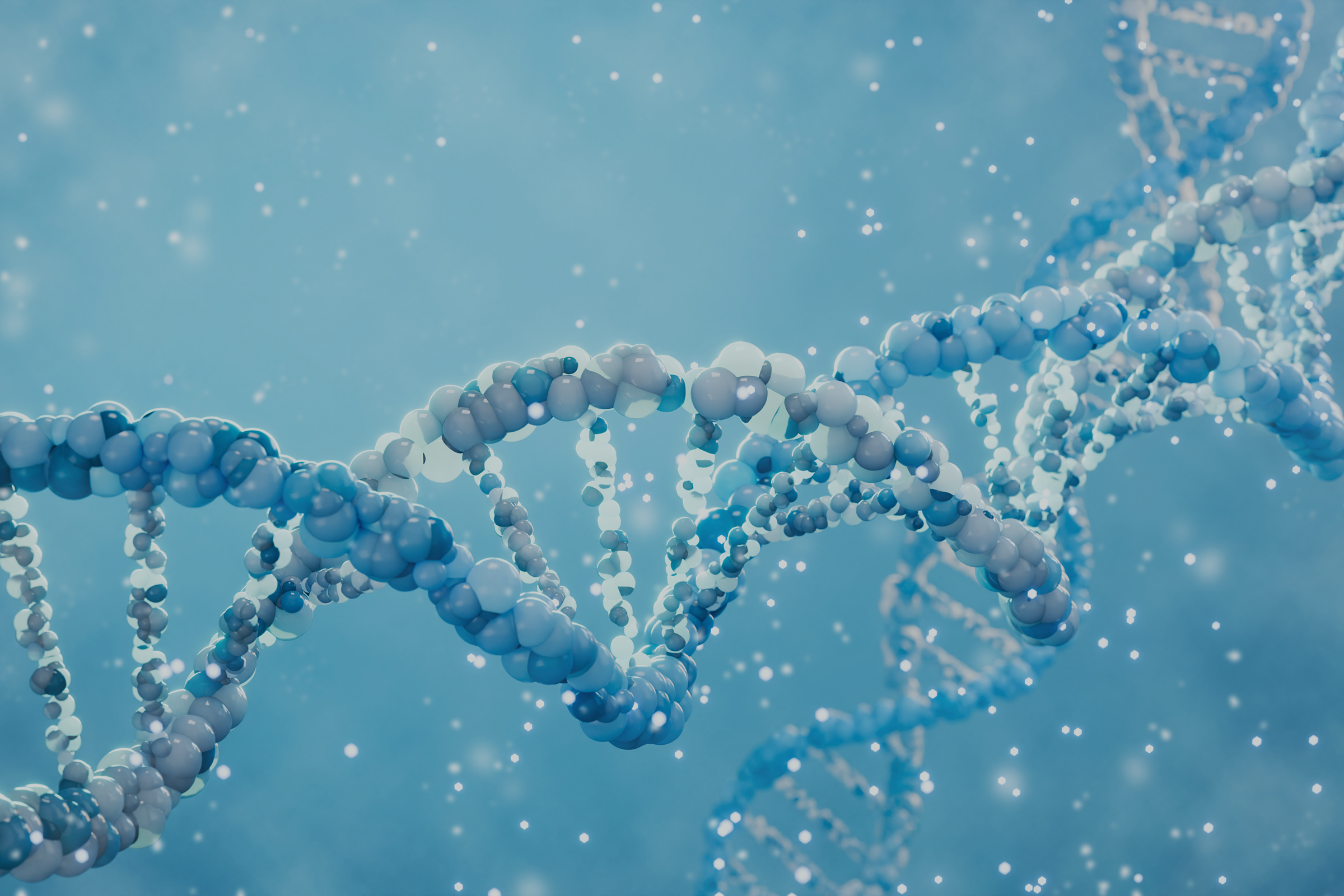
Nicotinamide adenine dinucleotide (NAD+) is a vital molecule required for energy production, DNA repair, and cellular maintenance across multiple hallmarks of aging. However, despite its essentiality, NAD+ levels are known to decline with age across multiple tissues.
Many of the hallmarks of aging are connected not just by accumulating damage, but by how cells use and lose NAD+ over time, making NAD+ decline a potential integrative node across the entire framework. There is a core feedback loop driving this connection: molecular damage activates NAD-consuming repair pathways, which reduces the cell’s ability to repair itself, which then generates more damage, and so on—a self-reinforcing cycle.
Key players here include poly(ADP-ribose) polymerases (PARPs), which respond to DNA damage by using NAD+ to help repair the genome,⁴ and CD38, an enzyme expressed in immune and other cells that degrades NAD+ as part of inflammatory signaling.⁵ Over time, this vicious cycle contributes to the progression of aging across multiple hallmarks.
In the following sections, we’ll walk through each hallmark, layer by layer, explaining what each is, how it drives aging, and where NAD+ plays a role in supporting cellular health.
Primary Hallmarks: Where Aging Begins at the Molecular Level
First, let’s take a look at primary hallmarks, which include genomic instability, telomere attrition, epigenetic alterations, and loss of proteostasis. These are the earliest forms of molecular damage and the roots from which all other hallmarks eventually emerge. These are the “wear and tear” processes that accumulate over time, setting the stage for downstream dysfunction across the other hallmarks. Supporting these processes is foundational to any aging intervention strategy, with the potential to reduce the burden of cellular damage, improve resilience, and influence multiple other hallmarks simultaneously.
Hallmark #1: Genomic Instability
DNA is the blueprint for every cell, guiding function, growth, and repair. However, DNA constantly faces damage—from internal sources like replication errors and reactive oxygen species (ROS),⁶ and external sources such as UV radiation, smoking, alcohol, environmental toxins,⁷ and even diet. Genomic instability refers to the accumulation of these chemical lesions over time.⁸ Healthy cells have repair systems in place to correct this damage, but these systems become less efficient with age, allowing lesions to compound.⁹
NAD+ plays a key role in maintaining genomic stability, as it’s a required substrate for PARPs, the enzymes that detect and repair DNA damage.¹⁰ When NAD+ levels decline, PARP activity is reduced, leaving DNA damage unrepaired. Preclinical studies show that restoring NAD+ levels enhances DNA repair capacity and supports cellular resilience.¹¹ Because genomic instability underlies many other hallmarks—including telomere attrition, mitochondrial dysfunction, and cellular senescence—it forms a foundation for aging. Unrepaired DNA can trigger senescence, disrupt epigenetic patterns, and accelerate broader decline.
Hallmark #2: Telomere Attrition
Telomere attrition, or shortening, is caused by many of the same factors that lead to DNA damage and genomic instability.¹² Telomeres are the protective structures at the ends of our chromosomes that can be likened to the plastic or metal endcaps on the tips of your shoelaces (known as aglets). These repetitive DNA sequences shorten with every cell division, protecting vital genetic information from being lost, just as aglets prevent shoelaces from fraying.
NAD+ helps maintain telomere integrity through sirtuins (SIRT1 and SIRT6), which regulate telomerase and support chromosome stability.¹³ As NAD+ declines with age, sirtuin activity drops, accelerating telomere shortening. Animal studies suggest that boosting NAD+—through nicotinamide riboside (NR) or CD38 inhibition¹⁴—can restore NAD+, improve mitochondrial function, and reduce aging-related phenotypes. While human research is still emerging, this highlights a potential pathway for supporting chromosomal health. Telomere attrition also contributes to epigenetic alterations, linking it to downstream hallmarks of aging.
Hallmark #3: Epigenetic Alterations
The epigenome is the system that controls which genes are turned on or off in each cell, guiding proper cellular function. Over time, or with repeated stress and environmental exposures, the epigenome becomes less precise—genes that should be silent may activate, and those that should be expressed may shut down. This gradual loss of epigenetic information contributes to aging and increases disease risk.¹⁵ Scientists track these changes with epigenetic clocks, which estimate biological age based on DNA methylation patterns.¹⁶ When a person’s biological age exceeds their chronological age, it signals accelerated aging at the cellular level, or epigenetic age acceleration (EAA).¹⁷
NAD+ plays a key role in the epigenome, as NAD-dependent enzymes such as SIRT1 and SIRT6 help maintain proper epigenetic patterns by regulating histone deacetylation and DNA methylation. As NAD+ drops, so does sirtuin activity, contributing to epigenetic dysregulation. Because epigenetic aging rates can vary widely from person to person, epigenetic clocks can help to identify who may benefit most from aging interventions.
Deep Dive: What Does the Latest Research Say About Nicotinamide Riboside, Exercise, and Epigenetic Aging?
A recent human study looked at whether NR supplementation and high-intensity interval training (HIIT) could slow epigenetic aging in skeletal muscle—a tissue particularly prone to age-related decline.¹⁸ Researchers measured biological age using seven epigenetic clocks across three separate interventions: adults took 1,000 mg of NR per day for five months, or completed either a four-week or six-week HIIT program.
They found that NR supplementation slowed biological aging in three of the seven clocks, including the MEAT clock (designed specifically for muscle), DunedinPACE, and PCHannum. However, one clock, PCGrimAge, showed a slight acceleration, highlighting that different clocks capture different aspects of epigenetic aging, and results need careful interpretation.
High-intensity exercise had almost the opposite effect, as the DunedinPACE clock showed a faster pace of aging after both HIIT interventions. Researchers note that this is likely due to a temporary cellular stress 48-72 hours after exercise, rather than true long-term aging acceleration. Interestingly, changes in the MEAT clock correlated with changes in mitochondrial content in both the NR and HIIT groups, suggesting that muscle-specific clocks are sensitive to mitochondrial health.
While this study focuses on NR in humans, early-stage lab research on related NAD+ precursors—such as nicotinamide mononucleotide (NMN)—has shown that NMN can similarly shift gene activity in human blood samples in ways associated with a younger biological age.¹⁹ These experiments were conducted ex vivo, meaning NMN was applied directly to the blood in a lab rather than taken orally.
Overall, this research demonstrates that epigenetic alterations—one of the foundational primary hallmarks—can be measurably influenced by NAD-boosting interventions, and that epigenetic clocks are emerging as valuable tools for tracking how our cells respond to lifestyle and supplementation strategies.
Hallmark #4: Loss of Proteostasis
Proteostasis, or protein homeostasis, keeps proteins properly folded and functional, which is essential for cellular health.²⁰ With age, protein quality control declines, allowing misfolded or damaged proteins to accumulate. This buildup contributes to diseases like Alzheimer’s, where tau and amyloid-beta plaques disrupt neurons,²¹ and Parkinson’s, where alpha-synuclein aggregates accumulate.²²
One defense against protein damage is autophagy—the cell’s internal recycling system that clears out dysfunctional proteins and other damaged molecules.²³ NAD+ supports this process, as it’s required for SIRT1-mediated activation of autophagy.²⁴ As NAD+ levels decline, autophagy slows, and protein damage accumulates, leading to deteriorated cellular health.
Boosting NAD+ in preclinical studies has improved autophagy²⁵ and reduced protein damage,²⁶ highlighting its role in maintaining cellular resilience. These primary hallmarks—genomic instability, telomere attrition, epigenetic alterations, and loss of proteostasis—represent the molecular origins of aging. Next, the body responds to this damage with protective mechanisms that are helpful initially but can become harmful over time—the antagonistic hallmarks.
Antagonistic Hallmarks: When the Body’s Own Defenses Become the Problem
After the primary hallmarks create molecular damage, the body activates its own set of protective responses. These antagonistic hallmarks—deregulated nutrient sensing, mitochondrial dysfunction, cellular senescence, and disabled macroautophagy—initially compensate for the damage, helping to maintain cellular and tissue function. However, when these responses persist over time, they shift from protective to harmful, driving further aging and representing a key inflection point in the aging process, as well as one of the most promising targets for interventions.
Hallmark #5: Deregulated Nutrient Sensing
Nutrient-sensing systems evolved to help us survive during periods of food scarcity.²⁷ When nutrients are abundant, cells can focus on growth, reproduction, and energy storage—their so-called “grow and build” mode, which activates pathways like mTOR and IIS (insulin and IGF-1 signaling).²⁷ But when nutrients are scarce, the body shifts to “repair and conserve” mode, activating pathways like AMPK²⁸ and sirtuins, which in turn trigger maintenance processes such as autophagy. In youth, these modes are finely balanced, but aging disrupts the homeostasis—IIS and mTOR can become chronically overactive, while AMPK and sirtuin activity declines, creating an environment that accelerates aging.
NAD+ plays a critical role in keeping the balance. Higher NAD+ levels activate sirtuins, which communicate with AMPK and inhibit mTOR,²⁹ essentially signaling to the cell to prioritize repair and maintenance (this is similar to the effects of caloric restriction). Preclinical studies have shown that boosting NAD+ in mice on a high-fat diet activated sirtuins, increased fat burning, and improved insulin sensitivity.³⁰ Blocking sirtuins eliminated these benefits, highlighting NAD+’s role in nutrient sensing and healthy aging. This shift in nutrient-sensing balance also affects the cell’s energy factories (the mitochondria), setting the stage for the next hallmark: mitochondrial dysfunction.
Hallmark #6: Mitochondrial Dysfunction
Mitochondria, the cell’s powerhouses, produce energy (ATP) but become less efficient with age, generating more reactive oxygen species (ROS) that damage mitochondrial DNA and worsen energy decline in a self-reinforcing cycle.³¹ NAD+ is essential for mitochondrial energy production, acting as an electron carrier in the electron transport chain.³² When NAD+ levels drop, energy production is reduced, leading to more ROS and mitochondrial dysfunction. Clinical studies show that boosting NAD+ can improve mitochondrial function,³³ stimulate mitochondrial biogenesis (the creation of new mitochondria),³⁴ and reduce oxidative stress.
Because mitochondria affect so many other hallmarks of aging, including genomic instability, loss of proteostasis, and epigenetic alterations, their dysfunction is one of the most consequential. This decline also sets the stage for the next hallmark, cellular senescence.
Hallmark #7: Cellular Senescence
Cellular senescence occurs when damaged or stressed cells permanently stop dividing.³⁵ These “zombie cells” remain metabolically active,³⁶ releasing pro-inflammatory molecules and growth factors—collectively called the senescence-associated secretory phenotype (SASP)—that can damage nearby tissue and drive aging-related disease. Senescence can be triggered by telomere shortening, DNA damage, or other stressors.
NAD+ is also part of this story, as senescent cells show elevated activity of enzymes like PARP and CD38,³⁷ which deplete NAD+ and further impair cellular function. In a study with aged mouse uteri, stromal cells exhibited both high senescence markers and low NAD+ levels.³⁸ Boosting NAD+ reduced senescence, restored cellular function, and improved tissue receptivity, highlighting its potential role in countering senescence-related decline.
Emerging therapies like senolytics aim to clear these harmful cells, and NAD+ support may enhance their effectiveness by helping remaining cells maintain proper function. This buildup of damaged cells also ties into the next hallmark—autophagy decline.
Hallmark #8: Disabled Macroautophagy
Macroautophagy, or autophagy, is the cell’s recycling system that clears damaged organelles, protein aggregates, and other cellular debris.³⁹ With age, this process slows, allowing dysfunctional mitochondria, misfolded proteins, and waste to build up, contributing to mitochondrial dysfunction, loss of proteostasis, and cellular senescence.
NAD+ is essential for autophagy because the NAD+-dependent enzyme SIRT1 activates autophagy-related genes.⁴⁰ Low NAD+ impairs this cleanup system, while boosting NAD+ enhances recycling and supports cellular health.⁴¹ Healthy autophagy is linked to longevity, showing that maintaining this protective process is critical.⁴² When autophagy and other antagonistic hallmarks fail, they set the stage for the integrative hallmarks—the visible effects of aging that we experience systemically.
Integrative Hallmarks: The Downstream Consequences That Drive Systemic Decline
Integrative hallmarks arise from the cumulative effects of both primary and antagonistic hallmarks. Over time, the molecular and cellular damage they create compounds, leading to system-wide dysfunction. These hallmarks are what we experience most directly as aging—the visible, functional changes that reduce quality of life and increase the risk of age-related diseases across multiple organ systems.
Hallmark #9: Stem Cell Exhaustion
Stem cells are undifferentiated cells (essentially blank canvases) that act as the body’s repair toolkit, having the ability to replace damaged or dying tissues. Most tissues rely on stem cell pools throughout life to maintain function or regenerate after injury. However, with age, stem cell populations decline in number and function, reducing the body’s ability to repair its tissues.⁴³ Cellular senescence, chronic inflammation from SASP, DNA damage, and telomere shortening all accelerate this exhaustion, contributing to organ dysfunction and disease.
NAD+ supports stem cell health by enabling sirtuins to maintain stem cell identity and self-renewal,⁴⁴ powering PARPs for DNA repair, and enabling mitophagy—a specialized type of autophagy specifically for clearing dysfunctional mitochondria. When NAD+ falls with age, all three mechanisms weaken, driving stem cell exhaustion. In aged mice, boosting NAD+ rejuvenated muscle stem cells and improved regenerative capacity, highlighting its key role in tissue maintenance.²⁶
Hallmark #10: Altered Intercellular Communication
Cells don’t operate in isolation; they are constantly sending and receiving messages and signals to one another to coordinate things like growth, repair, and immune responses. With age, these signaling networks become dysregulated, contributing to the chronic, low-grade inflammation that underlies most age-related diseases.
NAD+ plays a key role in keeping intercellular communication in check. NAD-dependent sirtuins suppress key inflammatory pathways,⁴⁵ including NF-κB, acting as a brake on excessive inflammation.⁴⁶ As NAD+ declines with age, this brake weakens, allowing chronic inflammation to rise. In turn, inflammation upregulates CD38, which depletes NAD+ and creates a damaging cycle of inflammation and NAD+ loss. Human studies show that NAD+ supplementation can modulate inflammatory markers,³³'⁴⁷ highlighting its potential to restore healthier cellular communication. This breakdown in communication not only drives inflammation but also feeds into other hallmarks, such as stem cell exhaustion, senescence, and mitochondrial dysfunction.
Hallmark #11: Chronic Inflammation (Inflammaging)
Chronic, low-grade inflammation (also known as “inflammaging”)⁴⁸ was recognized as its own hallmark of aging in 2023 because of its central role in aging and age-related disease.⁴⁹ Unlike acute inflammation, which is a short-term protective response to injury or infection, inflammaging persists over time, damaging tissues and organs throughout the body. Major drivers of inflammaging include many other hallmarks of aging, including cellular senescence (and SASP), mitochondrial dysfunction, microbial dysbiosis, and dysregulated intercellular signaling.
NAD+ is closely linked to inflammaging: as NAD+ levels fall with age, SIRT1 activity declines, releasing the brake on NF-κB-driven inflammatory gene expression and allowing chronic inflammation to take hold.⁵⁰ Human studies show that boosting NAD+ can reduce inflammatory markers,³³ highlighting its potential as part of a broader anti-inflammatory strategy. Because chronic inflammation contributes to nearly every age-related disorder, targeting inflammaging is considered one of the most promising and broadly applicable strategies for extending healthspan.
Hallmark #12: Dysbiosis
Gut dysbiosis was also added in 2023 as a newer hallmark of aging,² reflecting the growing recognition of the gut microbiome’s importance. This complex community of trillions of microbes in the digestive tract does more than support digestion—it also influences cognition, immunity, heart health, weight, and longevity.⁵¹ As we age, the microbiome shifts, often favoring less beneficial bacteria.⁵² This imbalance, known as dysbiosis, can contribute to increased intestinal permeability (“leaky gut”), systemic inflammation, and metabolic dysfunction.⁵³
Dysbiosis also affects NAD+ metabolism. The gut microbiome helps produce NAD+ precursors,⁵⁴ so age-related microbial changes reduce the body’s ability to maintain healthy NAD+ levels. Shifts in tryptophan metabolism with age further impair NAD+ availability.⁵⁴ In turn, lower NAD+ may worsen gut health, as preclinical studies suggest it supports intestinal barrier function and microbial balance.⁵⁵ Emerging research indicates that boosting NAD+ may help improve microbiome composition,³⁴ while supporting gut health through diet, fiber, and probiotics may enhance NAD+ metabolism—a bidirectional opportunity for intervention.
Hallmark #13: Extracellular Matrix Alterations
Extracellular matrix (ECM) alterations,⁵⁶ added in 2025 as one of the newest hallmarks of aging,³ reflect age-related changes in the structural network of proteins that support cells, tissue integrity, and repair.⁵⁷ With age, collagen and other components can fragment, stiffen, or cross-link excessively, impairing tissue flexibility and sometimes leading to fibrosis—scar tissue buildup that disrupts organ function.⁵⁸
Like several of the other hallmarks, NAD+ plays a role in ECM integrity through sirtuins,⁵⁹ which regulate antioxidant defense systems and anti-fibrotic pathways. As NAD+ declines with age, these protective mechanisms weaken, allowing abnormal ECM remodeling to accelerate. Preclinical research shows that increasing NAD+ can suppress the activation of scar-producing cells, reduce fibrotic proteins, and limit ECM accumulation in tissues like the liver.⁶⁰ ECM changes are also influenced by other hallmarks—like inflammation, senescence, and mitochondrial dysfunction—linking them to the broader aging network and setting the stage for the final hallmark.
Hallmark #14: Socio-Psychological Isolation
Socio-psychological isolation is the second newly-added hallmark of aging as of 2025,³ wisely reflecting that biological, psychological, and social factors are not separate entities but rather a deeply interconnected system that impacts health and aging. Prolonged isolation and stress are now known to chronically activate the body’s stress response system, the hypothalamic-pituitary-adrenal (HPA) axis.⁶¹ Over time, this suppresses an enzyme called NAMPT, which is responsible for producing NAD+.⁶² The resulting NAD+ depletion occurs in critical brain regions such as the prefrontal cortex and hippocampus, which govern mood, memory, and cognitive function.⁶³
In animal studies, this NAD+ loss weakens antioxidant defenses, disrupts mitochondrial function in neurons, and ultimately contributes to cellular damage linked to depressive symptoms and cognitive decline.⁶⁴ Restoring NAD+ reversed these stress-induced changes, improved mitochondrial function, and normalized behavior. This hallmark underscores the fact that healthy aging cannot be addressed through biology alone—social connection, stress management, and mental health are also measurable and vital to overall health and well-being.
Conclusion: NAD+ as a Central Hub: The Bigger Picture on Precision Modulation
Aging is more than just the passage of years; it also reflects the progressive disruption of balance across 14 interconnected biological and psycho-social processes. NAD+ sits at the heart of this network, acting as a central metabolic and signaling hub that touches every layer of the hallmarks of aging framework, from primary damage to antagonistic responses and integrative systemic decline. Because of this central role, NAD+ decline is increasingly seen not merely as a consequence of aging, but as a driver of the process itself.
The interconnected nature of the hallmarks means that supporting NAD+ metabolism may create beneficial ripple effects across multiple aging pathways. While this is promising, it is important to emphasize that NAD-boosting supplements are not a silver bullet, and the science is still evolving. However, preclinical and emerging human studies suggest that interventions targeting NAD+ precursors and oral supplementation may influence many hallmarks at once.
Ultimately, healthy aging is best approached as a multi-pronged strategy rather than a single intervention. Supporting NAD+ metabolism, alongside lifestyle, social, and environmental strategies, offers a promising path forward. As research continues to evolve, personalized strategies that integrate these insights may transform how we approach aging, shifting the focus from managing decline to actively promoting resilience and longevity.
References
- López-Otín, C., Blasco, M. A., Partridge, L., Serrano, M., & Kroemer, G. (2013). The Hallmarks of Aging. Cell, 153(6), 1194–1217. https://doi.org/10.1016/j.cell.2013.05.039
- López-Otín, C., Blasco, M. A., Partridge, L., Serrano, M., & Kroemer, G. (2023). Hallmarks of aging: An expanding universe. Cell, 186(2), 243–278. https://doi.org/10.1016/j.cell.2022.11.001
- López-Otín, C., & Kroemer, G. (2025). Hallmarks of aging: Integrating molecular and social determinants. Geromedicine. https://doi.org/10.70401/geromedicine.2025.0007
- Sousa, F. G., Matuo, R., Soares, D. G., Escargueil, A. E., Henriques, J. A. P., Larsen, A. K., & Saffi, J. (2012). PARPs and the DNA damage response. Carcinogenesis, 33(8), 1433–1440. https://doi.org/10.1093/carcin/bgs132
- Camacho-Pereira, J., Tarragó, M. G., Chini, C. C. S., Nin, V., Escande, C., Warner, G. M., Puranik, A. S., Schoon, R. A., Reid, J. M., Galina, A., & Chini, E. N. (2016). CD38 Dictates Age-Related NAD Decline and Mitochondrial Dysfunction through an SIRT3-Dependent Mechanism. Cell Metabolism, 23(6), 1127–1139. https://doi.org/10.1016/j.cmet.2016.05.006
- Chatterjee, N., & Walker, G. C. (2017). Mechanisms of DNA damage, repair, and mutagenesis. Environmental and Molecular Mutagenesis, 58(5), 235–263. https://doi.org/10.1002/em.22087
- Yousefzadeh, M., Henpita, C., Vyas, R., Soto-Palma, C., Robbins, P., & Niedernhofer, L. (2021). DNA damage—how and why we age? eLife, 10, e62852. https://doi.org/10.7554/elife.62852
- Vijg, J., & Suh, Y. (2013). Genome Instability and Aging. Physiology, 75(1), 645–668. https://doi.org/10.1146/annurev-physiol-030212-183715
- Gorbunova, V., Seluanov, A., Mao, Z., & Hine, C. (2007). Changes in DNA repair during aging. Nucleic Acids Research, 35(22), 7466–7474. https://doi.org/10.1093/nar/gkm756
- Murata, M. M., Kong, X., Moncada, E., Chen, Y., Imamura, H., Wang, P., Berns, M. W., Yokomori, K., & Digman, M. A. (2019). NAD+ consumption by PARP1 in response to DNA damage triggers metabolic shift critical for damaged cell survival. Molecular Biology of the Cell, 30(20), 2584–2597. https://doi.org/10.1091/mbc.e18-10-0650
- Fang, E. F., Kassahun, H., Croteau, D. L., Scheibye-Knudsen, M., Marosi, K., Lu, H., Shamanna, R. A., Kalyanasundaram, S., Bollineni, R. C., Wilson, M. A., Iser, W. B., Wollman, B. N., Morevati, M., Li, J., Kerr, J. S., Lu, Q., Waltz, T. B., Tian, J., Sinclair, D. A., … Bohr, V. A. (2016). NAD+ Replenishment Improves Lifespan and Healthspan in Ataxia Telangiectasia Models via Mitophagy and DNA Repair. Cell Metabolism, 24(4), 566–581. https://doi.org/10.1016/j.cmet.2016.09.004
- Shammas, M. A. (2011). Telomeres, lifestyle, cancer, and aging. Current Opinion in Clinical Nutrition and Metabolic Care, 14(1), 28–34. https://doi.org/10.1097/mco.0b013e32834121b1
- Amano, H., & Sahin, E. (2019). Telomeres and sirtuins: at the end we meet again. Molecular & Cellular Oncology, 6(5), e1632613. https://doi.org/10.1080/23723556.2019.1632613
- Sun, C., Wang, K., Stock, A. J., Gong, Y., Demarest, T. G., Yang, B., Giri, N., Harrington, L., Alter, B. P., Savage, S. A., Bohr, V. A., & Liu, Y. (2020). Re‐equilibration of imbalanced NAD metabolism ameliorates the impact of telomere dysfunction. The EMBO Journal, 39(21), e103420. https://doi.org/10.15252/embj.2019103420
- Gonzalo, S. (2010). Epigenetic alterations in aging. Journal of Applied Physiology, 109(2), 586–597. https://doi.org/10.1152/japplphysiol.00238.2010
- Teschendorff, A. E., & Horvath, S. (2025). Epigenetic ageing clocks: statistical methods and emerging computational challenges. Nature Reviews Genetics, 26(5), 350–368. https://doi.org/10.1038/s41576-024-00807-w
- Chervova, O., Panteleeva, K., Chernysheva, E., Widayati, T. A., Baronik, Ž. F., Hrbková, N., Schneider, J. L., Bobak, M., Beck, S., & Voloshin, V. (2024). Breaking new ground on human health and well-being with epigenetic clocks: A systematic review and meta-analysis of epigenetic age acceleration associations. Ageing Research Reviews, 102, 102552. https://doi.org/10.1016/j.arr.2024.102552
- Heikkinen, A., Uusitalo-Kylmälä, L., Blom, I., Helge, J. W., Gillberg, L., Seaborne, R., Larsen, S., Jacques, M., Grolaux, R., Aaltonen, S., Kaprio, J., Kolk, B. W. van der, Heinonen, S., Eynon, N., Pietiläinen, K. H., Kivelä, R., Pirinen, E., & Ollikainen, M. (2026). The divergent effects of nicotinamide riboside and high-intensity exercise training on skeletal muscle epigenetic aging. https://doi.org/10.64898/2026.01.16.26344093
- Shen, X., Wu, B., Jiang, W., Li, Y., Zhang, Y., Zhao, K., Nie, N., Gong, L., Liu, Y., Zou, X., Liu, J., Jin, J., & Ouyang, H. (2022). Scale bar of aging trajectories for screening personal rejuvenation treatments. Computational and Structural Biotechnology Journal, 20, 5750–5760. https://doi.org/10.1016/j.csbj.2022.10.021
- Labbadia, J., & Morimoto, R. I. (2014). The Biology of Proteostasis in Aging and Disease. Annual Review of Biochemistry, 84(1), 1–30. https://doi.org/10.1146/annurev-biochem-060614-033955
- Thapa, R., Bhat, A. A., Shahwan, M., Ali, H., PadmaPriya, G., Bansal, P., Rajotiya, S., Barwal, A., Prasad, G. V. S., Pramanik, A., Khan, A., Goh, B. H., Dureja, H., Singh, S. K., Dua, K., & Gupta, G. (2024). Proteostasis disruption and senescence in Alzheimer’s disease pathways to neurodegeneration. Brain Research, 1845, 149202. https://doi.org/10.1016/j.brainres.2024.149202
- Lehtonen, Š., Sonninen, T.-M., Wojciechowski, S., Goldsteins, G., & Koistinaho, J. (2019). Dysfunction of Cellular Proteostasis in Parkinson’s Disease. Frontiers in Neuroscience, 13, 457. https://doi.org/10.3389/fnins.2019.00457
- Glick, D., Barth, S., & Macleod, K. F. (2010). Autophagy: cellular and molecular mechanisms. The Journal of Pathology, 221(1), 3–12. https://doi.org/10.1002/path.2697
- Fang, E. F., Kassahun, H., Croteau, D. L., Scheibye-Knudsen, M., Marosi, K., Lu, H., Shamanna, R. A., Kalyanasundaram, S., Bollineni, R. C., Wilson, M. A., Iser, W. B., Wollman, B. N., Morevati, M., Li, J., Kerr, J. S., Lu, Q., Waltz, T. B., Tian, J., Sinclair, D. A., … Bohr, V. A. (2016). NAD+ Replenishment Improves Lifespan and Healthspan in Ataxia Telangiectasia Models via Mitophagy and DNA Repair. Cell Metabolism, 24(4), 566–581. https://doi.org/10.1016/j.cmet.2016.09.004
- Yuan, C., Yang, H., Lan, W., Yang, J., & Tang, Y. (2024). Nicotinamide ribose ameliorates myocardial ischemia/reperfusion injury by regulating autophagy and regulating oxidative stress. Experimental and Therapeutic Medicine, 27(5), 187. https://doi.org/10.3892/etm.2024.12475
- Zhang, H., Ryu, D., Wu, Y., Gariani, K., Wang, X., Luan, P., D’Amico, D., Ropelle, E. R., Lutolf, M. P., Aebersold, R., Schoonjans, K., Menzies, K. J., & Auwerx, J. (2016). NAD+ repletion improves mitochondrial and stem cell function and enhances life span in mice. Science, 352(6292), 1436–1443. https://doi.org/10.1126/science.aaf2693
- Efeyan, A., Comb, W. C., & Sabatini, D. M. (2015). Nutrient-sensing mechanisms and pathways. Nature, 517(7534), 302–310. https://doi.org/10.1038/nature14190
- Hardie, D. G., Ross, F. A., & Hawley, S. A. (2012). AMPK: a nutrient and energy sensor that maintains energy homeostasis. Nature Reviews Molecular Cell Biology, 13(4), 251–262. https://doi.org/10.1038/nrm3311
- Cantó, C., Gerhart-Hines, Z., Feige, J. N., Lagouge, M., Noriega, L., Milne, J. C., Elliott, P. J., Puigserver, P., & Auwerx, J. (2009). AMPK regulates energy expenditure by modulating NAD+ metabolism and SIRT1 activity. Nature, 458(7241), 1056–1060. https://doi.org/10.1038/nature07813
- Cantó, C., Houtkooper, R. H., Pirinen, E., Youn, D. Y., Oosterveer, M. H., Cen, Y., Fernandez-Marcos, P. J., Yamamoto, H., Andreux, P. A., Cettour-Rose, P., Gademann, K., Rinsch, C., Schoonjans, K., Sauve, A. A., & Auwerx, J. (2012). The NAD+ Precursor Nicotinamide Riboside Enhances Oxidative Metabolism and Protects against High-Fat Diet-Induced Obesity. Cell Metabolism, 15(6), 838–847. https://doi.org/10.1016/j.cmet.2012.04.022
- Murphy, M. P. (2009). How mitochondria produce reactive oxygen species. Biochemical Journal, 417(Pt 1), 1–13. https://doi.org/10.1042/bj20081386
- Stein, L. R., & Imai, S. (2012). The dynamic regulation of NAD metabolism in mitochondria. Trends in Endocrinology & Metabolism, 23(9), 420–428. https://doi.org/10.1016/j.tem.2012.06.005
- Zhou, B., Wang, D. D., Qiu, Y., Airhart, S., Liu, Y., Stempien-Otero, A., O’Brien, K. D., & Tian., R. (2020). Boosting NAD Level Suppresses Inflammatory Activation of PBMC in Heart Failure. Journal of Clinical Investigation, 130(11), 6054–6063. https://doi.org/10.1172/jci138538
- Lapatto, H. A. K., Kuusela, M., Heikkinen, A., Muniandy, M., Kolk, B. W. van der, Gopalakrishnan, S., Pöllänen, N., Sandvik, M., Schmidt, M. S., Heinonen, S., Saari, S., Kuula, J., Hakkarainen, A., Tampio, J., Saarinen, T., Taskinen, M.-R., Lundbom, N., Groop, P.-H., Tiirola, M., … Pirinen, E. (2023). Nicotinamide riboside improves muscle mitochondrial biogenesis, satellite cell differentiation, and gut microbiota in a twin study. Science Advances, 9(2), eadd5163. https://doi.org/10.1126/sciadv.add5163
- Huang, W., Hickson, L. J., Eirin, A., Kirkland, J. L., & Lerman, L. O. (2022). Cellular senescence: the good, the bad and the unknown. Nature Reviews. Nephrology, 18(10), 611–627. https://doi.org/10.1038/s41581-022-00601-z
- Nelson, B., & Faquin, W. (2023). Understanding the “zombie cells” that won’t die. Cancer Cytopathology, 131(10), 601–602. https://doi.org/10.1002/cncy.22765
- Chini, C. C. S., Cordeiro, H. S., Tran, N. L. K., & Chini, E. N. (2024). NAD metabolism: Role in senescence regulation and aging. Aging Cell, 23(1), e13920. https://doi.org/10.1111/acel.13920
- Hua, L., Liu, L., Gao, D., Ma, L., Jin, X., Lu, L., Tian, S., Jiang, X., Jin, C., Feng, B., Che, L., Xu, S., Lin, Y., Jin, L., Zhuo, Y., Li, M., & Wu, D. (2026). Myeloid‐Derived CD38 Mediates Age‐Related Endometrial Aging Through NAD+ Depletion. Aging Cell, 25(1). https://doi.org/10.1111/acel.70356
- Nieto-Torres, J. L., & Hansen, M. (2021). Macroautophagy and aging: The impact of cellular recycling on health and longevity. Molecular Aspects of Medicine, 82, 101020. https://doi.org/10.1016/j.mam.2021.101020
- Wilson, N., Kataura, T., Korsgen, M. E., Sun, C., Sarkar, S., & Korolchuk, V. I. (2023). The autophagy–NAD axis in longevity and disease. Trends in Cell Biology, 33(9), 788–802. https://doi.org/10.1016/j.tcb.2023.02.004
- Yuan, C., Yang, H., Lan, W., Yang, J., & Tang, Y. (2024). Nicotinamide ribose ameliorates myocardial ischemia/reperfusion injury by regulating autophagy and regulating oxidative stress. Experimental and Therapeutic Medicine, 27(5), 187. https://doi.org/10.3892/etm.2024.12475
- Nakamura, S., & Yoshimori, T. (2018). Autophagy and Longevity. Molecules and Cells, 41(1), 65–72. https://doi.org/10.14348/molcells.2018.2333
- Oh, J., Lee, Y. D., & Wagers, A. J. (2014). Stem cell aging: mechanisms, regulators and therapeutic opportunities. Nature Medicine, 20(8), 870–880. https://doi.org/10.1038/nm.3651
- Feng, Y., Qiu, H., & Chen, D. (2025). Regulation of Stem Cell Function by NAD+. Physiology, 40(4), 387–395. https://doi.org/10.1152/physiol.00052.2024
- Yang, Y., Liu, Y., Wang, Y., Chao, Y., Zhang, J., Jia, Y., Tie, J., & Hu, D. (2022). Regulation of SIRT1 and Its Roles in Inflammation. Frontiers in Immunology, 13, 831168. https://doi.org/10.3389/fimmu.2022.831168
- Liu, T., Zhang, L., Joo, D., & Sun, S.-C. (2017). NF-κB signaling in inflammation. Signal Transduction and Targeted Therapy, 2(1), 17023. https://doi.org/10.1038/sigtrans.2017.23
- Elhassan, Y. S., Kluckova, K., Fletcher, R. S., Schmidt, M. S., Garten, A., Doig, C. L., Cartwright, D. M., Oakey, L., Burley, C. V., Jenkinson, N., Wilson, M., Lucas, S. J. E., Akerman, I., Seabright, A., Lai, Y.-C., Tennant, D. A., Nightingale, P., Wallis, G. A., Manolopoulos, K. N., … Lavery, G. G. (2019). Nicotinamide Riboside Augments the Aged Human Skeletal Muscle NAD+ Metabolome and Induces Transcriptomic and Anti-inflammatory Signatures. Cell Reports, 28(7), 1717-1728.e6. https://doi.org/10.1016/j.celrep.2019.07.043
- Franceschi, C., Garagnani, P., Parini, P., Giuliani, C., & Santoro, A. (2018). Inflammaging: a new immune–metabolic viewpoint for age-related diseases. Nature Reviews Endocrinology, 14(10), 576–590. https://doi.org/10.1038/s41574-018-0059-4
- López-Otín, C., Blasco, M. A., Partridge, L., Serrano, M., & Kroemer, G. (2023). Hallmarks of aging: An expanding universe. Cell, 186(2), 243–278. https://doi.org/10.1016/j.cell.2022.11.001
- Schultz, M. B., Bochaton, T., Bonkowski, M., Li, J., Lokitiyakul, D., Colville, A., Gomes, A., & Sinclair, D. (2018). NAD+ DEPLETION AS A CAUSE OF INFLAMMAGING. Innovation in Aging, 2(suppl_1), 746–746. https://doi.org/10.1093/geroni/igy023.2751
- Vos, W. M. de, Tilg, H., Hul, M. V., & Cani, P. D. (2022). Gut microbiome and health: mechanistic insights. Gut, 71(5), 1020–1032. https://doi.org/10.1136/gutjnl-2021-326789
- Badal, V. D., Vaccariello, E. D., Murray, E. R., Yu, K. E., Knight, R., Jeste, D. V., & Nguyen, T. T. (2020). The Gut Microbiome, Aging, and Longevity: A Systematic Review. Nutrients, 12(12), 3759. https://doi.org/10.3390/nu12123759
- Acevedo-Román, A., Pagán-Zayas, N., Velázquez-Rivera, L. I., Torres-Ventura, A. C., & Godoy-Vitorino, F. (2024). Insights into Gut Dysbiosis: Inflammatory Diseases, Obesity, and Restoration Approaches. International Journal of Molecular Sciences, 25(17), 9715. https://doi.org/10.3390/ijms25179715
- Ren, Z., Xu, Y., Li, T., Sun, W., Tang, Z., Wang, Y., Zhou, K., Li, J., Ding, Q., Liang, K., Wu, L., Yin, Y., & Sun, Z. (2022). NAD+ and its possible role in gut microbiota: Insights on the mechanisms by which gut microbes influence host metabolism. Animal Nutrition, 10, 360–371. https://doi.org/10.1016/j.aninu.2022.06.009
- Novak, E. A., Crawford, E. C., Mentrup, H. L., Griffith, B. D., Fletcher, D. M., Flanagan, M. R., Schneider, C., Firek, B., Rogers, M. B., Morowitz, M. J., Piganelli, J. D., Wang, Q., & Mollen, K. P. (2023). Epithelial NAD+ depletion drives mitochondrial dysfunction and contributes to intestinal inflammation. Frontiers in Immunology, 14, 1231700. https://doi.org/10.3389/fimmu.2023.1231700
- McCabe, M. C., Hill, R. C., Calderone, K., Cui, Y., Yan, Y., Quan, T., Fisher, G. J., & Hansen, K. C. (2020). Alterations in extracellular matrix composition during aging and photoaging of the skin. Matrix Biology Plus, 8, 100041. https://doi.org/10.1016/j.mbplus.2020.100041
- Yue, B. (2014). Biology of the Extracellular Matrix. Journal of Glaucoma, 23(NA;), S20–S23. https://doi.org/10.1097/ijg.0000000000000108
- Zhou, H., Li, W., Pan, L., Zhu, T., Zhou, T., Xiao, E., & Wei, Q. (2024). Human extracellular matrix (ECM)-like collagen and its bioactivity. Regenerative Biomaterials, 11, rbae008. https://doi.org/10.1093/rb/rbae008
- Statzer, C., Park, J. Y. C., & Ewald, C. Y. (2023). Extracellular Matrix Dynamics as an Emerging yet Understudied Hallmark of Aging and Longevity. Aging and Disease, 14(3), 670–693. https://doi.org/10.14336/ad.2022.1116
- Zong, Z., Liu, J., Wang, N., Yang, C., Wang, Q., Zhang, W., Chen, Y., Liu, X., & Deng, H. (2021). Nicotinamide mononucleotide inhibits hepatic stellate cell activation to prevent liver fibrosis via promoting PGE2 degradation. Free Radical Biology and Medicine, 162, 571–581. https://doi.org/10.1016/j.freeradbiomed.2020.11.014
- Leistner, C., & Menke, A. (2020). Chapter 4 Hypothalamic–pituitary–adrenal axis and stress. Handbook of Clinical Neurology, 175, 55–64. https://doi.org/10.1016/b978-0-444-64123-6.00004-7
- Sun, X., Elangovan, V. R., Mapes, B., Camp, S. M., Sammani, S., Saadat, L., Ceco, E., Ma, S.-F., Flores, C., MacDougall, M. S., Quijada, H., Liu, B., Kempf, C. L., Wang, T., Chiang, E. T., & Garcia, J. G. N. (2014). The NAMPT Promoter Is Regulated by Mechanical Stress, Signal Transducer and Activator of Transcription 5, and Acute Respiratory Distress Syndrome–Associated Genetic Variants. American Journal of Respiratory Cell and Molecular Biology, 51(5), 660–667. https://doi.org/10.1165/rcmb.2014-0117oc
- Harrell, C. S., Gillespie, C. F., & Neigh, G. N. (2016). Energetic stress: The reciprocal relationship between energy availability and the stress response. Physiology & Behavior, 166, 43–55. https://doi.org/10.1016/j.physbeh.2015.10.009
- Xie, X., Xu, H., Shu, R., Du, S., Fan, H., Zhang, M., Sun, L., Zhou, J., Wang, L., Li, Z., & Anthony, D. C. (2025). Period3 modulates the NAD+-SIRT3 axis to alleviate depression-like behaviour by enhancing NAMPT activity in mice. Journal of Advanced Research, 77, 309–320. https://doi.org/10.1016/j.jare.2025.01.043