Can Increasing NAD+ Support Reproductive Health and Fertility?
Key Takeaways
- Reproductive aging is driven not only by time, but also by underlying changes in cellular energy production, mitochondrial function, and DNA repair.
- NAD+ is a central coenzyme in these processes that naturally declines with age in reproductive tissues, including the ovaries and testes.
- Preclinical research suggests that restoring NAD+ levels may improve egg quality, sperm function, and pregnancy-related outcomes by supporting cellular metabolism.
- Emerging human data indicate altered NAD+ metabolism in reproductive conditions, though clinical evidence is still limited.
- While NAD+ precursors such as NR and NMN are being actively studied, their role in fertility and pregnancy in humans has not been fully established.
Many people assume that declining fertility is simply an inevitable consequence of reaching their late 30s and 40s, but in reality, the story starts much earlier at the cellular level. Long before changes like irregular periods or missed ovulation appear, reproduction places substantial demands on the human body, requiring continuous energy to grow eggs and sperm, support hormone production, build and remodel the uterine environment, and sustain a developing pregnancy.¹ All of these processes depend on healthy mitochondria,² intact DNA repair systems,³ and coordinated stress responses that must remain functional for years, often decades, before someone ever tries to conceive.
As world-leading NAD+ researcher Dr. Charles Brenner and others have emphasized, reproduction can be viewed as a kind of “stress test” for metabolism—indeed, as Dr. Brenner puts it, “postpartum is the mother of metabolic stressors”—revealing vulnerabilities in cellular energy systems that have accumulated with age, environmental exposures, and lifestyle factors.
While age is an undisputed driver of reduced reproductive capacity, it often reflects deeper changes in cellular function, including metabolism, mitochondrial health, and DNA repair pathways. At the center of this network is nicotinamide adenine dinucleotide (NAD+), a molecule found in every cell that helps convert nutrients into energy, supports mitochondrial function, and fuels enzymes involved in DNA repair, epigenetic regulation, and cellular stress responses.
NAD+ levels naturally decline with age in many tissues—including the ovaries⁴ and testes⁵—and emerging research suggests that this decline may be an underappreciated piece of the fertility puzzle, potentially contributing to changes in egg quality, ovarian reserve, sperm function, and pregnancy outcomes. Both scientific researchers and online patient communities have shown growing interest in NAD+ and reproductive health, reflecting a broader curiosity about targeting NAD+ stores for fertility. However, well‑controlled human studies are still needed to determine if and how these approaches translate into meaningful clinical benefits.
In this article, we’ll dive deeper into the biology of reproductive aging—across both female and male systems—and why it begins earlier than most people realize. We’ll then examine how NAD+ fits into this picture, focusing on its role in cellular energy, genomic stability, and mitochondrial function, before walking through the current state of research on NAD+ and reproductive health, including ovarian aging and egg quality, sperm health, conception, in vitro fertilization (IVF), prenatal and postpartum health, and potential long-term effects on offspring.
Reproductive Health and the Biology of Reproductive Aging
Reproductive health is about more than just the ability to conceive; it also encompasses hormonal balance, egg or sperm quality, ovarian reserve, and the body’s capacity to support implantation, pregnancy, and a healthy birth. This coordination of multiple biological systems is tightly interconnected, and all are influenced by increasing age.
The ovaries are widely recognized as one of the earliest organs in the body to exhibit signs of biological aging—often decades before other systems show obvious changes.⁶ This means that egg quality and quantity decline over time, even in people who are otherwise healthy. While ovarian reserve (the number of remaining follicles) declines with age,⁷ it is the integrity of the egg itself—its mitochondrial function, chromosomal stability, and ability to properly divide—that plays a more critical role in successful conception and healthy embryonic development.⁸
However, reproductive aging does not stop at the ovaries. Over time, hormonal patterns can shift, the uterine environment can alter in ways that can influence implantation or placental development, and cellular damage accumulates across reproductive tissues. Across years and decades, these changes can impact cycle regularity, implantation rates, pregnancy outcomes, and responses to fertility treatments like IVF.
Despite what many believe, male reproductive health is also impacted by aging (although it’s more gradual and often later in life).⁹ Sperm count, motility, and morphology can all decline, while DNA fragmentation and other measures of genomic integrity tend to worsen over time.⁹ These changes can influence time to conceive, miscarriage risk, and the health of resulting embryos, which is why male reproductive aging is increasingly recognized as a meaningful piece of the fertility puzzle.
Together, all of these age-related changes—across the ovaries, uterus, hormones, and sperm—share the fact that they are driven (at least in part) by what is happening inside the cell.
NAD+ and the Cellular Basis of Reproductive Aging
NAD+ is a coenzyme found in every cell in the body, where it plays a vital role in energy production, DNA repair, and mitochondrial function. However, as we age, NAD+ levels are known to decline across multiple tissues, which can impair mitochondrial efficiency, disrupt normal cellular signaling, and weaken genomic stability. These changes are especially relevant for reproductive cells, as they require high energy needs and tightly controlled cell division.¹⁰
Egg and sperm cells are among the most energy-dependent cells in the body, relying heavily on NAD+ and healthy mitochondria for processes like proper maturation, division, and development.¹⁰ This makes egg and sperm cells especially vulnerable to NAD+ decline, resulting in compromised cellular processes required for successful fertilization and embryonic development.¹¹
Because oocytes are formed before birth and then maintained for decades, they may be particularly susceptible to drops in NAD+ and the effects of oxidative stress and DNA damage over time. Emerging research suggests that the ovary, which is already one of the earliest organs to show signs of aging, also shows changes in NAD+ metabolism that parallel declines in ovarian reserve and oocyte quality.¹¹
Studies have also connected altered NAD+ pathways with reproductive health disorders like polycystic ovarian syndrome (PCOS), including disruptions in NAD-related enzymes and metabolites, indicating local metabolic stress within the ovary.¹² A recent study showed altered NAD+ metabolism in women with recurrent miscarriage, suggesting that changes in the NAD+ salvage pathway may be linked to adverse pregnancy outcomes.¹³ While these findings reflect that NAD+ metabolism is disrupted, not necessarily deficient, they still highlight how NAD-related pathways may be involved in reproductive health.
A key player in this connection is CD38, an enzyme that breaks down NAD+ and whose activity increases with age.¹⁴ In animal models, CD38 expression has been shown to rise in the ovaries over time, contributing to NAD+ decline and reduced fertility in mice.¹⁵ This suggests that age-related increases in NAD+ consumption may be one mechanistic link between declining metabolic and reproductive health.
Data from human ovarian tissues have shown similar results. In a study with IVF patients, nicotinamide levels in follicular fluid were significantly lower in women with markers of ovarian aging, alongside increases in its metabolite 1-methylnicotinamide (MNA).¹⁶ This ratio was strongly associated with age and negatively correlated with measures of ovarian reserve, including antral follicle count, anti-Mullerian hormone (AMH) levels, and the number of oocytes retrieved. These findings suggest that shifts in NAD+ metabolism may serve as a measurable indicator of ovarian aging in humans.
Given its central role in energy production and DNA repair, NAD+ is uniquely positioned to support the immense metabolic demands of reproduction, fertility, and pregnancy. Cells involved in reproduction—particularly oocytes and sperm—depend heavily on healthy mitochondrial function, making them especially sensitive to disruptions in NAD+ availability. NAD+ levels can be supported through precursors like nicotinamide riboside (NR) and nicotinamide mononucleotide (NMN), both of which have been investigated in the context of aging and, increasingly, reproductive health.¹⁷
NAD+ Across the Female Reproductive Lifespan
A woman’s reproductive journey spans decades, from the years of active fertility, including conception and pregnancy, to the postpartum period and the long-term health of her children. NAD+ plays a role at every one of these stages, supporting the cellular processes required for successful reproduction and development.
Because NAD+ declines with age, this reduction may intersect with reproductive challenges at several points along the trajectory—not just during the narrow “fertility window.” In this section, we’ll follow the research through that natural progression, beginning with egg quality and conception, then moving through pregnancy and lactation, and finally considering how maternal NAD+ status might influence the health of the next generation.
Although most of the studies so far are preclinical, the consistency of findings across multiple stages of reproduction makes NAD+ an especially compelling focus for ongoing research. Plus, emerging human data—such as the changes in NAD+ metabolism observed in follicular fluid¹⁶—suggest that these patterns may extend beyond animal models and into clinical relevance.
Fertility, Egg Quality, and Ovarian Health
Female fertility is fundamentally tied to the health of the ovaries and the eggs they produce, and both are acutely sensitive to age-related cellular decline. Oocytes are among the most mitochondria-rich cells in the body,¹⁸ making them highly dependent on NAD+ and reflecting the substantial energy required to support their maturation, fertilization, and early embryonic development.
As NAD+ levels drop, mitochondrial efficiency declines, leading to increased oxidative stress and disruptions in cellular processes that are critical for maintaining egg quality.¹¹ Over time, this cascade can contribute to chromosomal abnormalities, impaired embryo development, and the reduced likelihood of successful conception.
Across multiple preclinical studies, low NAD+ has been associated with ovarian aging, while restoration of NAD+ has been shown to improve key fertility outcomes.¹⁹ For example, in aged mice, boosting ovarian NAD+ with precursors like NR²⁰ and NMN²¹ led to improved mitochondrial function,²² increased ovulation rates, and higher live-birth rates,²⁰ suggesting that NAD+ availability may play an important role in supporting oocyte quality and reproductive function.
This relationship is not only seen with natural age-related fertility decline, but also in metabolic and reproductive conditions. For example, in PCOS, studies suggest that age-related pathways are disrupted in ways that increase oxidative stress and are linked with poorer egg quality.²³ In diabetes and insulin resistance, high blood sugar and chronic inflammation place additional stress on ovarian cells, further straining NAD+ levels and mitochondrial function.²⁴
Together, these findings suggest that NAD+ plays a central role in maintaining ovarian function and egg quality, and that declines in NAD+ may be a key mechanism linking both aging and metabolic dysfunction to reduced fertility.
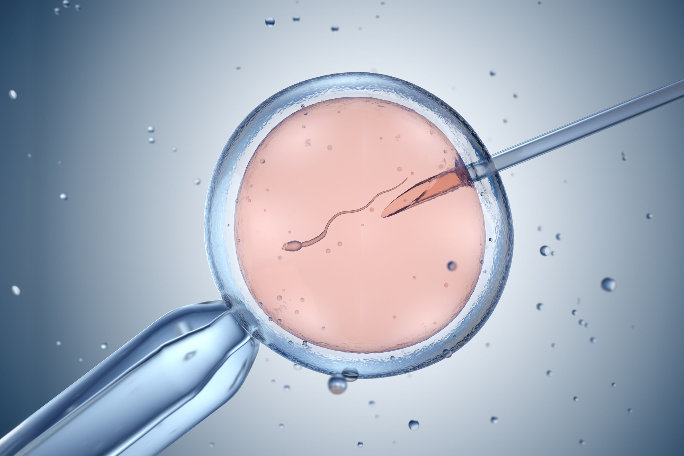
Conception & In Vitro Fertilization (IVF)
For many individuals and couples, conception may ultimately require medical support, with IVF being one of the most widely used and consequential fertility treatments. But even with modern lab techniques, a persistent challenge with IVF remains: embryo arrest, a state in which many embryos stop developing before they reach a viable stage.²⁵
In many cases, the underlying “why” of embryo arrest remains unknown—but recent research has begun to shed light on the biology behind it. Arrested embryos often show signs of cellular energy deficits and a quiescent- or senescent‑like state, with disrupted metabolism and stress responses that appear to stop normal development.²⁶ Given NAD+’s role in cellular energy, researchers have suggested that supporting NAD+-related pathways may improve embryo survival in the IVF setting.
Building on this idea, researchers have started to test how supporting NAD+ pathways during assisted reproduction might influence outcomes.²⁶ For example, experimental work with adding NAD+ precursors to maturation or culture media has reported improvements in oocyte maturation and embryo development in IVF-like animal models,¹⁷'²⁷ suggesting that better cellular energy status may translate into more embryos reaching later stages.
Although these findings are encouraging, they remain early-stage and preclinical, and well‑controlled clinical studies in humans are still needed before any NAD-targeted approaches can be recommended or integrated into routine IVF practice. Beyond implantation and early development, NAD+ may also play a role in supporting pregnancy itself through its influence on placental and fetal energy metabolism.
Pregnancy & Maternal Health
Pregnancy is a period of extraordinary metabolic demand, as the body must simultaneously support maternal physiology, placental development, and fetal growth—all of which require substantial cellular energy. NAD+ is central to meeting these demands, and emerging preclinical research has connected depleted NAD+ stores to a range of adverse outcomes, from placental dysfunction²⁸ to fetal growth impairment.²⁹
Across several models, low NAD+ levels or disrupted NAD+ metabolism have been linked to pregnancy challenges. In a study looking at the placentas from women with inflammation‑driven preeclampsia, as well as an animal model, placental NAD+ was found to be depleted, while boosting NAD+ with NR improved placental mitochondrial function, reduced oxidative stress and inflammation, and helped to prevent maternal hypertension and fetal growth restriction.²⁸
Other preclinical research has examined pregnancy stressors like gestational hypoglycemia,³⁰ gestational hypoxia,²⁹ and drug exposures,³¹ and similarly found that disrupted NAD+ metabolism is associated with worse maternal-fetal outcomes, while NAD+ restoration partially normalized oxidative stress, placental function, and fetal growth.
These studies consistently tell the same story: when NAD+ is depleted, or NAD+ metabolism is disrupted during pregnancy, the placenta and fetus are more vulnerable to metabolic stress—and that restoring NAD+ can at least partially improve these outcomes.
Postpartum & Lactation
While pregnancy and even pre-conception are often talked about widely in health conversations, the postpartum period is often overlooked, despite placing some of the highest metabolic demands on the body—especially if a woman is lactating or breastfeeding.
Breast milk production is an energetically intensive process, significantly increasing daily energy expenditure. Emerging research has suggested that NAD+ metabolism may play a meaningful role in both milk quality and maternal and infant health. In a rodent model, maternal NR supplementation during lactation increased nursing behaviors, restored low postpartum NAD+ levels, enhanced milk production, supported maternal metabolic health, and improved offspring growth and neurodevelopment.³² Similarly, NR supplementation in postpartum sows led to increased NAD+ in breast milk, boosted milk yield, improved milk nutrient composition, and produced heavier, healthier piglets at weaning.³³
Although neither of these preclinical models represents human data, these findings help to build a mechanistic case that replenishing NAD+ during the postpartum period may support the intense energetic demands of lactation and may influence both maternal and infant health outcomes.
Offspring Development
Scientists increasingly use the term “developmental programming” to describe how conditions in the womb and early life can leave lasting biological imprints on a child’s health, shaping metabolism, cognition, and disease risk later in life. If maternal NAD+ status potentially helps to define the cellular environment in which a fetus develops and is nourished, this raises the question of whether its effects might extend well beyond birth, into childhood and adulthood.
Many of the preclinical studies mentioned in previous sections have explored this idea. In models where maternal NAD+ is depleted—whether by stress, inflammation, or altered placental function—their offspring are more likely to show signs of impaired growth,³³ altered brain development,³² and changes in metabolism³² that persist after birth. Conversely, when NAD+ is restored, offspring show partial or significant recovery in these areas. Supplementary visual data from these studies further illustrate how shifts in maternal NAD+ status may manifest across multiple physiological systems.
Overall, these studies suggest that maternal NAD+ stores may do more than support just day-to-day cellular energy needs; they may also influence long-term offspring health. However, as these are preclinical models, more research is needed with human studies to see if similar patterns hold true with people.
NAD+ and Male Reproductive Health
While fertility conversations often center on women, male factor infertility contributes to an estimated 30-50% of cases, making male reproductive health an equally important part of the picture. Like oocytes, sperm rely heavily on healthy mitochondrial function—and, therefore, also depend on NAD+ to generate the energy required for motility, as well as to support DNA repair processes that preserve sperm integrity.
In preclinical research, low NAD+ levels have been linked to reduced sperm count, lower motility, increased DNA damage, and impaired sperm cell development. When testicular NAD+ was depleted in young male mice, spermatogenesis declined, sperm counts fell to levels seen in aged animals, and the testes became smaller³⁴ and structurally disorganized.³⁵ In one study with mice, restoring NAD+ with NR³⁶ reversed these spermatogenic defects and reduced the associated DNA damage.³⁷
Beyond age, environmental and genetic factors that disturb NAD+ metabolism can also compromise male reproductive health. Heat stress, a common environmental challenge for the testes, has been shown in a boar model to lower testicular NAD+ and worsen semen quality.³⁸ In the same model, supplementation with NR restored NAD+, improved semen quality, and reduced oxidative and inflammatory testes damage.³⁸
Taken together, these findings support the emerging view that age‑related NAD+ decline in men may help link metabolic aging, declining sperm quality, and intergenerational health outcomes, while also underscoring how limited direct human data still are in this area.⁵
The Honest Caveats: What Science Still Doesn’t Know
Although mostly preclinical, the studies described in this article paint a compelling picture of how NAD+ plays a role in reproductive health and aging—but we must be clear about what the science can and cannot currently support. The overwhelming majority of the evidence comes from animal models and ex vivo systems, and translation of these findings to humans is not guaranteed.
The most human-relevant data point—the widely cited work on arrested IVF embryos—used human embryos to show that many arrested embryos enter a quiescent- or senescent‑like state and that metabolic interventions like NR can coax some of them back into development.²⁶ However, the NR portion of the study was carried out in culture and was not part of a randomized clinical trial in patients, so it cannot tell us whether supplementing people undergoing IVF will actually improve live birth rates.
There are also several question marks on the population level, including the fact that the overall prevalence of NAD+ deficiency in pregnant women is not yet known.³⁹ Plus, while the preclinical evidence remains consistent and promising for the use of NAD+ precursors like NR and NMN, questions remain about the optimal dosing, timing, and safety of these supplements during preconception, pregnancy, and lactation.
Several clinical trials are now underway or recently completed that will hopefully shed some light on these unknowns. In lactating women, the MOONRISE trial is testing whether oral NR can increase milk volume in mothers of extremely preterm infants. Regarding fertility, the ENHANCE trial and a head‑to‑head comparison of NR versus vitamin E are studying the effects of NR on egg quality or IVF outcomes in women of advanced maternal age.
Similarly, NMN-focused trials are studying the effects on embryo development after prior IVF failure, diminished ovarian reserve, or PCOS. The results of these studies will be critical in determining whether NAD+ precursors can move from promising preclinical tools to evidence‑based options in human reproductive care.
Conclusion: A Promising Frontier Worth Watching
NAD+ sits at the center of the cellular responses that power reproduction, including mitochondrial energy production, DNA repair, and epigenetic regulation. NAD+’s age-related decline intersects meaningfully with reproductive aging in both women and men. Across stages of fertility, pregnancy, postpartum or lactation, and offspring development, a consistent body of preclinical evidence has emerged suggesting that supporting NAD+ with precursors like NR and NMN may counteract certain aspects of reproductive decline in animal models.
At the same time, while early human data points to disrupted NAD+ metabolism in certain reproductive situations, most of the evidence still remains preclinical at this time. For now, the most clearly actionable steps include lifestyle factors that support healthy NAD+ balance—such as regular exercise, balanced nutrition, sufficient sleep, and stress management—which are broadly beneficial for metabolic and reproductive health regardless of supplementation.
NAD+ represents a promising and rapidly evolving area of fertility science, which may ultimately expand the toolkit for supporting reproductive health across the lifespan. That said, until more robust human data emerges, informed decision-making guided by emerging research and clinical expertise remains essential for anyone considering NAD‑targeted strategies in the context of fertility and pregnancy.
References
- Fontana, R., & Torre, S. (2016). The Deep Correlation between Energy Metabolism and Reproduction: A View on the Effects of Nutrition for Women Fertility. Nutrients, 8(2), 87. https://doi.org/10.3390/nu8020087
- Rasool, A., Muneeb, J. M., Thakur, S., & Manzoor, I. (2026). Beyond the Powerhouse – Unveiling Hidden Influence of Mitochondria on Reproductive Health and Fertility. Fertility Science and Research, 13, 2. https://doi.org/10.25259/fsr_51_2025
- Titus, S., Li, F., Stobezki, R., Akula, K., Unsal, E., Jeong, K., Dickler, M., Robson, M., Moy, F., Goswami, S., & Oktay, K. (2013). Impairment of BRCA1-Related DNA Double-Strand Break Repair Leads to Ovarian Aging in Mice and Humans. Science Translational Medicine, 5(172), 172ra21. https://doi.org/10.1126/scitranslmed.3004925
- Liang, J., Huang, F., Song, Z., Tang, R., Zhang, P., & Chen, R. (2023). Impact of NAD+ metabolism on ovarian aging. Immunity & Ageing, 20(1), 70. https://doi.org/10.1186/s12979-023-00398-w
- Feuz, M. B., Nelson, D. C., Miller, L. B., Zwerdling, A. E., Meyer, R. G., & Meyer-Ficca, M. L. (2024). Current Insights & a Potential Role of NAD in the Reproductive Health of Aging Fathers and Their Children. Reproduction (Cambridge, England), 167(6), e230486. https://doi.org/10.1530/rep-23-0486
- Wang, G., Yang, R., & Zhang, H. (2025). Ovarian vascular aging: a hidden driver of mid-age female fertility decline. NPJ Aging, 11(1), 24. https://doi.org/10.1038/s41514-025-00216-1
- Amanvermez, R., & Tosun, M. (2016). An Update on Ovarian Aging and Ovarian Reserve Tests. International Journal of Fertility & Sterility, 9(4), 411–415. https://doi.org/10.22074/ijfs.2015.4591
- Hirano, M., Onodera, T., Takasaki, K., Takahashi, Y., Ichinose, T., Nishida, H., Hiraike, H., & Nagasaka, K. (2025). Ovarian aging: pathophysiology and recent developments in maintaining ovarian reserve. Frontiers in Endocrinology, 16, 1619516. https://doi.org/10.3389/fendo.2025.1619516
- Kumar, N., Singh, A. K., & Choudhari, A. R. (2017). Impact of age on semen parameters in male partners of infertile couples in a rural tertiary care center of central India: A cross-sectional study. International Journal of Reproductive BioMedicine, 15(8), 497–502. https://doi.org/10.29252/ijrm.15.8.497
- Martin, W. (2017). Oocytes, Maternal Information and Functions. Results and Problems in Cell Differentiation, 63, 373–387. https://doi.org/10.1007/978-3-319-60855-6_16
- Cordone, V., Vergara, T., Falone, S., Tatone, C., & Emidio, G. D. (2025). Is NAD+ a key factor in ovarian aging and dysfunction? Insights and uncertainties from current research. Biology of Reproduction, 114(2), 463–478. https://doi.org/10.1093/biolre/ioaf140
- Ahmed, M., Riaz, U., Lv, H., & Yang, L. (2024). A Molecular Perspective and Role of NAD+ in Ovarian Aging. International Journal of Molecular Sciences, 25(9), 4680. https://doi.org/10.3390/ijms25094680
- Cuny, H., Shand, A. W., Goth, J., Sheng, D. Z., Tossey, T., Martin, E. M. M. A., Sipka, A., Aleshin, O., Schneuer, F. J., Nassar, N., & Dunwoodie, S. L. (2025). Identification of potential NAD-related biomarkers of recurrent miscarriage risk. Human Reproduction, deaf195. https://doi.org/10.1093/humrep/deaf195
- Hogan, K. A., Chini, C. C. S., & Chini, E. N. (2019). The Multi-faceted Ecto-enzyme CD38: Roles in Immunomodulation, Cancer, Aging, and Metabolic Diseases. Frontiers in Immunology, 10(1187), 1187. https://doi.org/10.3389/fimmu.2019.01187
- Perrone, R., Kumaar, P. V. A., Haky, L., Hahn, C., Riley, R., Balough, J., Zaza, G., Soygur, B., Hung, K., Prado, L., Kasler, H. G., Tiwari, R., Matsui, H., Hormazabal, G. V., Heckenbach, I., Scheibye-Knudsen, M., Duncan, F. E., & Verdin, E. (2023). CD38 regulates ovarian function and fecundity via NAD+ metabolism. iScience, 26(10), 107949. https://doi.org/10.1016/j.isci.2023.107949
- Bocca, C., Boguenet, M., Boucret, L., Bouet, P., Reynier, P., & May‐Panloup, P. (2026). Nicotinamide (Vitamin B3) Deficiency in Follicular Fluid of Patients With Ovarian Ageing. Journal of Cellular and Molecular Medicine, 30(6). https://doi.org/10.1111/jcmm.71085
- Bertoldo, M. J., Listijono, D. R., Ho, W.-H. J., Riepsamen, A. H., Goss, D. M., Richani, D., Jin, X. L., Mahbub, S., Campbell, J. M., Habibalahi, A., Loh, W.-G. N., Youngson, N. A., Maniam, J., Wong, A. S. A., Selesniemi, K., Bustamante, S., Li, C., Zhao, Y., Marinova, M. B., … Wu, L. E. (2020). NAD+ Repletion Rescues Female Fertility during Reproductive Aging. Cell Reports, 30(6), 1670-1681.e7. https://doi.org/10.1016/j.celrep.2020.01.058
- Zhang, D., Keilty, D., Zhang, Z. F., & Chian, R. C. (2017). Mitochondria in oocyte aging: current understanding. Facts, Views & Vision in ObGyn, 9(1), 29–38.
- Yang, Q., Li, H., Wang, H., Chen, W., Zeng, X., Luo, X., Xu, J., & Sun, Y. (2023). Deletion of enzymes for de novo NAD+ biosynthesis accelerated ovarian aging. Aging Cell, 22(9), e13904. https://doi.org/10.1111/acel.13904
- Yang, Q., Cong, L., Wang, Y., Luo, X., Li, H., Wang, H., Zhu, J., Dai, S., Jin, H., Yao, G., Shi, S., Hsueh, A. J., & Sun, Y. (2020). Increasing ovarian NAD+ levels improve mitochondrial functions and reverse ovarian aging. Free Radical Biology and Medicine, 156, 1–10. https://doi.org/10.1016/j.freeradbiomed.2020.05.003
- Liang, J., Huang, F., Hao, X., Zhang, P., & Chen, R. (2024). Nicotinamide mononucleotide supplementation rescues mitochondrial and energy metabolism functions and ameliorates inflammatory states in the ovaries of aging mice. MedComm, 5(10), e727. https://doi.org/10.1002/mco2.727
- Arslan, N. P., Akpinar, Z., Aybek, H., Doymus, M., Asilkan-Kaldik, G., Esim, N., & Taskin, M. (2025). NAD+ precursors mitigate the in vitro and in vivo reproductive defects: Limitations and possible solutions. Reproductive Toxicology, 138, 109067. https://doi.org/10.1016/j.reprotox.2025.109067
- Zhu, Z., Lei, M., Guo, R., Xu, Y., Zhao, Y., Wei, C., Yang, Q., & Sun, Y. (2025). Nicotinamide riboside supplementation ameliorates ovarian dysfunction in a PCOS mouse model. Journal of Ovarian Research, 18(1), 9. https://doi.org/10.1186/s13048-025-01596-4
- Wei, C., Zeng, X., Wang, K., Wang, M., Lei, M., Zhu, Z., Xu, Y., Zhao, Y., Yang, Q., & Sun, Y. (2025). Nicotinamide riboside supplementation protects against maternal diabetes-associated decline in oocyte quality. Reproduction (Cambridge, England), 169(5), e240350. https://doi.org/10.1530/rep-24-0350
- Sahin, G. N., Yildirim, R. M., & Seli, E. (2023). Embryonic arrest: causes and implications. Current Opinion in Obstetrics and Gynecology, 35(3), 184–192. https://doi.org/10.1097/gco.0000000000000871
- Yang, Y., Shi, L., Fu, X., Ma, G., Yang, Z., Li, Y., Zhou, Y., Yuan, L., Xia, Y., Zhong, X., Yin, P., Sun, L., Zhang, W., Babarinde, I. A., Wang, Y., Zhao, X., Hutchins, A. P., & Tong, G. (2022). Metabolic and epigenetic dysfunctions underlie the arrest of in vitro fertilized human embryos in a senescent-like state. PLoS Biology, 20(6), e3001682. https://doi.org/10.1371/journal.pbio.3001682
- POLLARD, C.-L., GIBB, Z., HAWDON, A., SWEGEN, A., & GRUPEN, C. G. (2021). Supplementing media with NAD+ precursors enhances the in vitro maturation of porcine oocytes. The Journal of Reproduction and Development, 67(5), 319–326. https://doi.org/10.1262/jrd.2021-080
- Jahan, F., Vasam, G., Cariaco, Y., Nik-Akhtar, A., Green, A., Menzies, K. J., & Bainbridge, S. A. (2024). NAD+ depletion is central to placental dysfunction in an inflammatory subclass of preeclampsia. Life Science Alliance, 7(12), e202302505. https://doi.org/10.26508/lsa.202302505
- Thompson, L. P., Song, H., & Hartnett, J. (2023). Nicotinamide Riboside, an NAD + Precursor, Protects Against Cardiac Mitochondrial Dysfunction in Fetal Guinea Pigs Exposed to Gestational Hypoxia. Reproductive Sciences, 1–12. https://doi.org/10.1007/s43032-023-01387-6
- Lee, S. R., Jeong, S. H., Mukae, M., Kim, S.-Y., Ko, J.-W., Kwun, H.-J., & Hong, E.-J. (2023). Dietary supplementation with nicotinamide riboside improves fetal growth under hypoglycemia. The Journal of Nutritional Biochemistry, 116, 109310. https://doi.org/10.1016/j.jnutbio.2023.109310
- Liu, F., He, J., Chen, X., Liu, R., Li, F., Geng, Y., Dai, Y., Zhang, Y., Wang, Y., & Mu, X. (2024). Maternal Administration of Acetaminophen Affects Meiosis Through its Metabolite NAPQI Targeting SIRT7 in Fetal Oocytes. Antioxidants & Redox Signaling, 41(1–3), 93–109. https://doi.org/10.1089/ars.2023.0270
- Ear, P. H., Chadda, A., Gumusoglu, S. B., Schmidt, M. S., Vogeler, S., Malicoat, J., Kadel, J., Moore, M. M., Migaud, M. E., Stevens, H. E., & Brenner, C. (2019). Maternal Nicotinamide Riboside Enhances Postpartum Weight Loss, Juvenile Offspring Development, and Neurogenesis of Adult Offspring. Cell Reports, 26(4), 969-983.e4. https://doi.org/10.1016/j.celrep.2019.01.007
- Huang, L., Yang, X., Pan, C., Zhang, W., Li, Y., Zhang, R., Li, H., Li, Y., Zhuo, Y., Jiang, X., Che, L., Lin, Y., Xu, S., Fang, Z., Feng, B., Wu, D., & Hua, L. (2026). Effects of nicotinamide riboside supplementation during late gestation and lactation on sow performance, milk metabolome, and gut microbiome. Journal of Animal Science and Biotechnology, 17(1), 26. https://doi.org/10.1186/s40104-025-01339-x
- Meyer-Ficca, M. L., Zwerdling, A. E., Swanson, C. A., Tucker, A. G., Lopez, S. A., Wandersee, M. K., Warner, G. M., Thompson, K. L., Chini, C. C. S., Chen, H., Chini, E. N., & Meyer, R. G. (2022). Low NAD+ Levels Are Associated With a Decline of Spermatogenesis in Transgenic ANDY and Aging Mice. Frontiers in Endocrinology, 13, 896356. https://doi.org/10.3389/fendo.2022.896356
- Ni, F., Wang, F., Li, J., Liu, Y., Sun, X., Chen, J., Li, J., Zhang, Y., Jin, J., Ye, X., Tu, M., Chen, J., Chen, C., & Zhang, D. (2023). BNC1 deficiency induces mitochondrial dysfunction-triggered spermatogonia apoptosis through the CREB/SIRT1/FOXO3 pathway: the therapeutic potential of nicotinamide riboside and metformin. Biology of Reproduction, 110(3), 615–631. https://doi.org/10.1093/biolre/ioad168
- Xu, Y., Wang, H., Li, H., Wei, C., Zhu, Z., Zhao, Y., Zhu, J., Lei, M., Sun, Y., & Yang, Q. (2025). Nicotinamide Riboside Supplementation Alleviates Testicular Aging Induced by Disruption of Qprt‐Dependent NAD+ De Novo Synthesis in Mice. Aging Cell, 24(6), e70004. https://doi.org/10.1111/acel.70004
- Li, C., Bi, R., Wang, L., Ma, Y.-H., Yao, Y.-G., Zheng, P., China, S. K. L. of G. R. and E., Kunming Institute of Zoology, Chinese Academy of Sciences, Kunming, Yunnan 650201, China, K. L. of A. M. and H. D. M. of Y. P., Kunming Institute of Zoology, Chinese Academy of Sciences, Kunming, Yunnan 650201, China, K. C. of L. S., University of Chinese Academy of Sciences, Kunming, Yunnan 650204, China, N. R. C. for N.-H. P., National Research Facility for Phenotypic &. Genetic Analysis of Model Animals (Primate Facility), Kunming Institute of Zoology, Chinese Academy of Sciences, Kunming, Yunnan 650107, & China, K. J. L. of B. and M. R. in C. D., Kunming Institute of Zoology, Chinese Academy of Sciences, Kunming, Yunnan 650204,. (2023). Characterization of long-term ex vivo expansion of tree shrew spermatogonial stem cells. Zoological Research, 44(6), 1080–1094. https://doi.org/10.24272/j.issn.2095-8137.2023.317
- Jin, X., Luo, X., Shen, W., Lv, G., Jiang, X., Jin, C., Feng, B., Che, L., Xu, S., Lin, Y., Zhuo, Y., Wu, D., & Hua, L. (2026). Nicotinamide Riboside Alleviates Heat Stress‐Induced Intestinal Dysfunction by Enhancing Antioxidant Capacity, Restoring Immune Homeostasis, and Modulating Gut Microbiota in a Boar Model. Molecular Nutrition & Food Research, 70(5), e70418. https://doi.org/10.1002/mnfr.70418
- Dunwoodie, S. L., Bozon, K., Szot, J. O., & Cuny, H. (2023). Nicotinamide Adenine Dinucleotide Deficiency and Its Impact on Mammalian Development. Antioxidants & Redox Signaling, 39(16), 1108–1132. https://doi.org/10.1089/ars.2023.0349