Article: What Is Mitochondrial Health? Energy, NAD+, and the Science of Aging
What Is Mitochondrial Health? Energy, NAD+, and the Science of Aging
Key Takeaways
- Mitochondria sit at the center of aging. Mitochondrial health is central to how the body produces energy, responds to stress, and moves through the aging process.
- When mitochondria falter, risk goes up. Dysfunctional mitochondria produce less energy and more oxidative stress and inflammation, increasing vulnerability to a range of age-related diseases.
- NAD+ is essential for mitochondria. NAD+ is required for mitochondrial energy production and key cellular repair processes, and its levels decline substantially with age.
- NAD+ precursors show early promise. Human studies of niacin, NR, and NMN suggest that restoring NAD+ can improve certain markers of mitochondrial capacity, biogenesis, or stress handling, although the evidence is still evolving.
- Mitochondrial support is a long-term, integrative strategy. The most grounded way to support mitochondrial health is through lasting lifestyle habits, potentially complemented by NAD+ precursors.
Although many people experience fatigue, weakness, and physical deterioration as they grow older, these changes are not inevitable consequences of aging. As research on aging and longevity progresses, it’s becoming clear that these outcomes result from breakdowns in molecular processes occurring deep within our cells. What was previously thought to be an unavoidable part of aging is now being viewed as a result of dysfunctional cellular machinery—especially in our mitochondria, the energy powerhouses that are considered regulators of cellular health and metabolism.¹
The health of our mitochondria is central to the conversation about aging, with their health and resilience recognized as among the most significant determinants of how we age and how resilient our bodies remain over time.² Influencing everything from metabolism and inflammation to DNA repair and stress responses, mitochondrial health plays a crucial role in longevity and overall well-being.³ In this article, we’ll dive deeper into what mitochondria do, how they decline—and what that means for long-term health—and a look into what the current evidence says about supporting their function with age.
What Is Mitochondrial Health and Why Does It Matter?
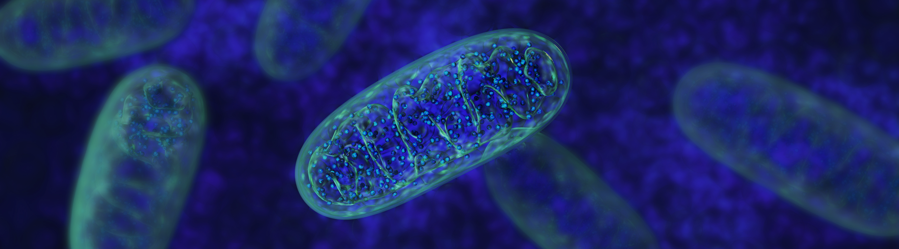
Mitochondria are microscopic, double-membraned organelles found in nearly every cell in the human body, serving as the primary site of cellular energy production, converting the food we eat into ATP (adenosine triphosphate). ATP fuels nearly every action our bodies perform, from muscle contraction to neuronal signaling to wound healing.⁴
But mitochondria are more than just energy generators—they also regulate metabolism, balance oxidative stress, and maintain cellular quality control.³ When functioning optimally, they generate adequate ATP with minimal production of reactive oxygen species (ROS). Conversely, dysfunctional mitochondria produce less energy and generate more oxidative stress, leading to negative downstream consequences for nearly every organ and system in the body.⁵
However, mitochondrial health is not a fixed state—their health and function are dynamic, continuously shaped by age, lifestyle, nutrition, environmental exposures, or disease.⁶ Our cells constantly monitor mitochondrial performance through quality control systems that can identify and remove damaged organelles while generating new, healthy ones (a process called mitochondrial biogenesis).⁷ When these systems work properly, cells maintain a healthy balance of functional mitochondria capable of fueling physical activity, efficiently processing nutrients, and protecting against chronic disease and age-related physiological decline.
How Mitochondria Produce Energy and Keep the Body Functioning
Mitochondria are responsible for producing the vast majority of our cellular energy—roughly 90% of the ATP that cells need to survive and function optimally. Through highly efficient biochemical processes, mitochondria provide a steady supply of ATP to cells, enabling them to maintain both their most basic and complex functions.¹ Some of the body’s organs/tissues are highly metabolically active—like the heart, brain, and skeletal muscles—meaning they rely heavily on mitochondrial output. The heart beats approximately 100,000 times per day,⁸ the brain processes vast amounts of information while accounting for roughly 20% of the body's total energy expenditure,⁹ and skeletal muscles power everyday movement and maintain posture, making these organs/tissues particularly dependent on consistent, efficient mitochondrial performance. When mitochondrial function falters, consequences can become apparent quickly,¹⁰ including fatigue, cognitive dysfunction, reduced physical endurance, or cardiovascular strain.¹¹
Mitophagy: How Cells Recycle Damaged Mitochondria
Some of the main functions of mitochondria include regulating inflammatory signaling,¹² coordinating cellular repair processes,¹³ and facilitating mitophagy,¹⁴ the selective removal of damaged or dysfunctional mitochondria. Mitophagy acts as a quality-control system, as cells identify mitochondria that are no longer working well, tag them for breakdown, and recycle their components so new, healthy mitochondria can take their place.
When this process is working efficiently, it helps maintain a population of high-functioning mitochondria that can keep up with the cell’s energy demands and stressors over time. Experimental research suggests that NAD+ availability is one of the factors that help to keep this system running, with NAD+ repletion shown to restore impaired mitophagy and improve mitochondrial quality in aging and disease models.¹⁵
But when mitophagy and other quality control mechanisms decline, damaged mitochondria can accumulate, producing excessive ROS and inflammatory signals that accelerate aging and disease.¹⁶ Therefore, mitochondrial health influences not just how much energy a cell can generate, but also how well cells maintain resilience and resist age-related decline.
What Is Mitochondrial Dysfunction? How It Accelerates Aging and Disease
Mitochondrial dysfunction—the progressive loss of mitochondrial efficiency and quantity—is recognized as one of the hallmarks of aging.¹⁷ Over time, cells generate fewer new mitochondria, while existing ones accumulate damage and become less capable of producing ATP efficiently.¹⁸ This simultaneous decline in quantity and quality means aging cells have fewer mitochondrial resources, and those that remain can become increasingly impaired.
Mitochondrial dysfunction can have far-reaching implications, starting with diminished energy availability that limits tissues’ ability to maintain normal function.¹⁷ Cellular repair processes can slow, and mitochondria become less efficient at managing oxidative stress, allowing ROS to build up and damage cellular components. Damaged mitochondria also release more inflammatory signals that activate immune pathways and contribute to chronic inflammation or “inflammaging.”¹⁹
From there, mitochondrial damage can spill out into age-related conditions, with links to neurodegeneration,²⁰ metabolic disorders including type 2 diabetes,²¹ cardiovascular disease, and sarcopenia.²² Now, many researchers understand mitochondrial decline to not just be a downstream consequence of aging, but a driving mechanism of it, actively accelerating the aging process and creating a self-reinforcing feedback loop in which dysfunction begets further cellular damage.
What Is NAD+ and How Does It Support Mitochondrial Health?
Nicotinamide adenine dinucleotide (NAD+) is a coenzyme found in every cell in the body, where it supports a wide range of critical functions—including helping mitochondria efficiently convert nutrients into ATP.²³ During energy production, NAD+ acts as a molecular carrier, shuttling electrons so mitochondria can run the reactions that generate ATP and supply the continuous energy needs of tissues like the heart, brain, and skeletal muscle.²⁴
However, NAD+ levels decline substantially with age,²⁵ and this drop tends to run in parallel with the reduced mitochondrial function observed in older tissues.¹⁸ Lower NAD+ levels can impair mitochondrial energy production and weaken the activity of NAD-dependent enzymes that support cellular health, like sirtuins and PARPs.²⁶ As a result, declining NAD+ is increasingly viewed as one of the molecular links between aging, mitochondrial dysfunction, and reduced cellular resilience.
Therefore, restoring NAD+ levels has emerged as a promising strategy to support mitochondrial health—and, with it, healthy aging and overall well-being.²⁷ As we’ll dive into more in the next section, studies show that elevating NAD+ is associated with improvements in mitochondrial quality and quantity,²⁸ suggesting that NAD+ availability may play an important role in how well mitochondria function over the lifespan.
NAD+ levels can be influenced by lifestyle and nutritional strategies, including regular physical activity,²⁹ caloric restriction,³⁰ and the intake of NAD+ precursors³¹—compounds such as nicotinamide mononucleotide (NMN) and nicotinamide riboside (NR) that convert into NAD+ in the body. Together, these approaches help form a toolkit that researchers are actively studying to understand how best to sustain NAD+ and mitochondrial health with age.
NAD+ and Mitochondrial Health: What Human Clinical Studies Reveal
For years, much of what was known about NAD+ and mitochondrial health came from cell-based³² and animal³³ studies, which consistently suggested that restoring NAD+ can improve mitochondrial function. However, while this type of research is essential for understanding mechanisms, human studies are needed to determine whether these strategies have real-world relevance for consumers and clinicians in the context of aging and disease prevention.
Early clinical research is beginning to offer a clearer picture of what NAD+ repletion might mean. In adults living with mitochondrial myopathy—a genetic condition that causes muscle dysfunction due to defective mitochondria—high-dose niacin (an NAD+ precursor) not only increased NAD+ levels in blood and muscle tissue but was also associated with improvements in muscle strength and performance, along with increased markers of mitochondrial biogenesis, the process by which new mitochondria are formed.³⁴ In people with heart failure—a condition where systemic inflammation and oxidative stress are common—supplementation with NR was found to elevate blood NAD+ while also reducing inflammation and improving mitochondrial respiration in white blood cells.³⁵ Older, sedentary men have shown similar patterns, with niacin supplementation leading to better measures of mitochondrial content in skeletal muscle and mitochondrial respiration.³⁶ Taken together, these studies suggest that, in several high-energy-demand tissues, NAD+ precursor supplementation may support a stronger and more resilient mitochondrial network.
Additional human studies with NR have reported improvements in markers of mitochondrial health in certain populations. In a twin study, NR supplementation was associated with increased mitochondrial biogenesis and enhanced muscle stem cell differentiation,²⁸ suggesting that NAD+ repletion may influence how muscle tissues remodel and maintain regenerative capacity. Trials in people with chronic conditions, such as chronic kidney disease, have also hinted that NR may improve mitochondrial function and reduce oxidative and inflammatory stress in specific immune cells.³⁷
However, a recent study that combined NMN with blood-flow-restricted exercise—a training method where cuffs are used to partially restrict blood flow to working muscles during low-load exercise—was more nuanced.³⁸ The researchers found that while NMN appeared to reduce inflammatory signaling in skeletal muscle after exercise, it also blunted the usual exercise-induced gains in mitochondrial content. This suggests that, while NAD+ repletion may help mitigate inflammation, it may also, in some contexts, interfere with the adaptive stress responses that exercise is meant to trigger. Rather than undermining the link between NAD+ and mitochondria, this research highlights the complexity of NAD+ biology and the importance of context, dosing, and timing when considering interventions.
The overall direction of the human evidence so far points toward a meaningful relationship between NAD+ and mitochondria, with studies consistently showing that NAD+ repletion improves some measures of mitochondrial capacity, biogenesis, or stress handling, especially in metabolically stressed or older populations. At the same time, the field is still developing, and more research is needed to clarify who might benefit most and which precursors and doses are optimal.
Mitochondrial Health, NAD+, and Longevity: Key Insights and Practical Implications
Overall, the growing science around mitochondrial health paints a clear picture: how well our mitochondria function is a foundational pillar of how the body produces energy, responds to stress, and moves through the aging process. Healthy, well-regulated, and efficient mitochondria support physical and cognitive performance, metabolic health, and resilience across organs that demand consistent energy, from the heart to the brain to the skeletal muscles. When mitochondrial function declines, the result is not only less energy, but also more oxidative stress, inflammation, and vulnerability to age-related disease.
NAD+ has emerged as a promising avenue for supporting mitochondrial function, with early human studies with NR, NMN, and niacin suggesting that replenishing NAD+ can improve at least some markers of mitochondrial capacity, biogenesis, or stress handling in specific tissues and populations. While the research evolves, the most grounded way to approach mitochondrial support is as a long-term, integrative strategy rooted in lifestyle habits like regular physical activity and healthy nutrition, informed by evolving clinical evidence, and, where appropriate, complemented by targeted NAD+ precursors. As our understanding of cellular health continues to deepen, mitochondrial function—and the NAD-dependent processes that sustain it—is increasingly recognized as a central component of how we age and how we feel doing so.
References
- Casanova, A., Wevers, A., Navarro-Ledesma, S., & Pruimboom, L. (2023). Mitochondria: It is all about energy. Frontiers in Physiology, 14, 1114231. https://doi.org/10.3389/fphys.2023.1114231
- Harrington, J. S., Ryter, S. W., Plataki, M., Price, D. R., & Choi, A. M. K. (2023). Mitochondria in health, disease, and aging. Physiological Reviews, 103(4), 2349–2422. https://doi.org/10.1152/physrev.00058.2021
- Javadov, S., Kozlov, A. V., & Camara, A. K. S. (2020). Mitochondria in Health and Diseases. Cells, 9(5), 1177. https://doi.org/10.3390/cells9051177
- McBride, H. M., Neuspiel, M., & Wasiak, S. (2006). Mitochondria: More Than Just a Powerhouse. Current Biology, 16(14), R551–R560. https://doi.org/10.1016/j.cub.2006.06.054
- Xu, X., Pang, Y., & Fan, X. (2025). Mitochondria in oxidative stress, inflammation and aging: from mechanisms to therapeutic advances. Signal Transduction and Targeted Therapy, 10(1), 190. https://doi.org/10.1038/s41392-025-02253-4
- Akbari, M., Kirkwood, T. B. L., & Bohr, V. A. (2019). Mitochondria in the signaling pathways that control longevity and health span. Ageing Research Reviews, 54, 100940–100940. https://doi.org/10.1016/j.arr.2019.100940
- Popov, L. (2020). Mitochondrial biogenesis: An update. Journal of Cellular and Molecular Medicine, 24(9), 4892–4899. https://doi.org/10.1111/jcmm.15194
- Shaffer, F., McCraty, R., & Zerr, C. L. (2014). A healthy heart is not a metronome: an integrative review of the heart’s anatomy and heart rate variability. Frontiers in Psychology, 5, 1040. https://doi.org/10.3389/fpsyg.2014.01040
- Balasubramanian, V. (2021). Brain power. Proceedings of the National Academy of Sciences, 118(32), e2107022118. https://doi.org/10.1073/pnas.2107022118
- Filler, K., Lyon, D., Bennett, J., McCain, N., Elswick, R., Lukkahatai, N., & Saligan, L. N. (2014). Association of mitochondrial dysfunction and fatigue: A review of the literature. BBA Clinical, 1, 12–23. https://doi.org/10.1016/j.bbacli.2014.04.001
- Zhou, B., & Tian, R. (2018). Mitochondrial dysfunction in pathophysiology of heart failure. Journal of Clinical Investigation, 128(9), 3716–3726. https://doi.org/10.1172/jci120849
- Marchi, S., Guilbaud, E., Tait, S. W. G., Yamazaki, T., & Galluzzi, L. (2023). Mitochondrial control of inflammation. Nature Reviews Immunology, 23(3), 159–173. https://doi.org/10.1038/s41577-022-00760-x
- Osellame, L. D., Blacker, T. S., & Duchen, M. R. (2012). Cellular and molecular mechanisms of mitochondrial function. Best Practice & Research Clinical Endocrinology & Metabolism, 26(6), 711–723. https://doi.org/10.1016/j.beem.2012.05.003
- Li, A., Gao, M., Liu, B., Qin, Y., chen, L., Liu, H., Wu, H., & Gong, G. (2022). Mitochondrial autophagy: molecular mechanisms and implications for cardiovascular disease. Cell Death & Disease, 13(5), 444. https://doi.org/10.1038/s41419-022-04906-6
- Fang, E. F., Hou, Y., Lautrup, S., Jensen, M. B., Yang, B., SenGupta, T., Caponio, D., Khezri, R., Demarest, T. G., Aman, Y., Figueroa, D., Morevati, M., Lee, H.-J., Kato, H., Kassahun, H., Lee, J.-H., Filippelli, D., Okur, M. N., Mangerich, A., … Bohr, V. A. (2019). NAD+ augmentation restores mitophagy and limits accelerated aging in Werner syndrome. Nature Communications, 10(1), 5284. https://doi.org/10.1038/s41467-019-13172-8
- Murphy, M. P. (2009). How mitochondria produce reactive oxygen species. Biochemical Journal, 417(Pt 1), 1–13. https://doi.org/10.1042/bj20081386
- Zong, Y., Li, H., Liao, P., Chen, L., Pan, Y., Zheng, Y., Zhang, C., Liu, D., Zheng, M., & Gao, J. (2024). Mitochondrial dysfunction: mechanisms and advances in therapy. Signal Transduction and Targeted Therapy, 9(1), 124. https://doi.org/10.1038/s41392-024-01839-8
- Chistiakov, D. A., Sobenin, I. A., Revin, V. V., Orekhov, A. N., & Bobryshev, Y. V. (2014). Mitochondrial Aging and Age‐Related Dysfunction of Mitochondria. BioMed Research International, 2014(1), 238463. https://doi.org/10.1155/2014/238463
- Fulop, T., Larbi, A., Pawelec, G., Khalil, A., Cohen, A. A., Hirokawa, K., Witkowski, J. M., & Franceschi, C. (2023). Immunology of Aging: the Birth of Inflammaging. Clinical Reviews in Allergy & Immunology, 64(2), 109–122. https://doi.org/10.1007/s12016-021-08899-6
- Klemmensen, M. M., Borrowman, S. H., Pearce, C., Pyles, B., & Chandra, B. (2024). Mitochondrial dysfunction in neurodegenerative disorders. Neurotherapeutics, 21(1), e00292. https://doi.org/10.1016/j.neurot.2023.10.002
- Prasun, P. (2020). Mitochondrial dysfunction in metabolic syndrome. Biochimica et Biophysica Acta (BBA) - Molecular Basis of Disease, 1866(10), 165838. https://doi.org/10.1016/j.bbadis.2020.165838
- Marzetti, E., Calvani, R., Cesari, M., Buford, T. W., Lorenzi, M., Behnke, B. J., & Leeuwenburgh, C. (2013). Mitochondrial dysfunction and sarcopenia of aging: From signaling pathways to clinical trials. The International Journal of Biochemistry & Cell Biology, 45(10), 2288–2301. https://doi.org/10.1016/j.biocel.2013.06.024
- Covarrubias, A. J., Perrone, R., Grozio, A., & Verdin, E. (2021). NAD+ metabolism and its roles in cellular processes during ageing. Nature Reviews Molecular Cell Biology, 22(2), 119–141. https://doi.org/10.1038/s41580-020-00313-x
- Cantó, C., Menzies, K. J., & Auwerx, J. (2015). NAD+ Metabolism and the Control of Energy Homeostasis: A Balancing Act between Mitochondria and the Nucleus. Cell Metabolism, 22(1), 31–53. https://doi.org/10.1016/j.cmet.2015.05.023
- McReynolds, M. R., Chellappa, K., & Baur, J. A. (2020). Age-related NAD+ decline. Experimental Gerontology, 134, 110888. https://doi.org/10.1016/j.exger.2020.110888
- Yusri, K., Jose, S., Vermeulen, K. S., Tan, T. C. M., & Sorrentino, V. (2025). The role of NAD+ metabolism and its modulation of mitochondria in aging and disease. Npj Metabolic Health and Disease, 3(1), 26. https://doi.org/10.1038/s44324-025-00067-0
- Sun, Y., Han, J.-C., Tran, K., Pham, T., Zhou, Q., & Lu, J. (2026). An integrated anti-aging framework targeting NAD+ homeostasis, mitochondrial quality control, and redox stability: Roles of NMN/NR, PQQ, and EGT. Redox Biology, 93, 104191. https://doi.org/10.1016/j.redox.2026.104191
- Lapatto, H. A. K., Kuusela, M., Heikkinen, A., Muniandy, M., Kolk, B. W. van der, Gopalakrishnan, S., Pöllänen, N., Sandvik, M., Schmidt, M. S., Heinonen, S., Saari, S., Kuula, J., Hakkarainen, A., Tampio, J., Saarinen, T., Taskinen, M.-R., Lundbom, N., Groop, P.-H., Tiirola, M., … Pirinen, E. (2023). Nicotinamide riboside improves muscle mitochondrial biogenesis, satellite cell differentiation, and gut microbiota in a twin study. Science Advances, 9(2), eadd5163. https://doi.org/10.1126/sciadv.add5163
- Walzik, D., Jonas, W., Joisten, N., Belen, S., Wüst, R. C. I., Guillemin, G., & Zimmer, P. (2023). Tissue‐specific effects of exercise as NAD+‐boosting strategy: Current knowledge and future perspectives. Acta Physiologica, e13921. https://doi.org/10.1111/apha.13921
- Poljsak, B., Kovač, V., & Milisav, I. (2020). Healthy Lifestyle Recommendations: Do the Beneficial Effects Originate from NAD+ Amount at the Cellular Level? Oxidative Medicine and Cellular Longevity, 2020, 8819627. https://doi.org/10.1155/2020/8819627
- Yaku, K., & Nakagawa, T. (2023). NAD+ Precursors in Human Health and Disease: Current Status and Future Prospects. Antioxidants & Redox Signaling, 39(16), 1133–1149. https://doi.org/10.1089/ars.2023.0354
- Katsyuba, E., Mottis, A., Zietak, M., Franco, F. D., Velpen, V. van der, Gariani, K., Ryu, D., Cialabrini, L., Matilainen, O., Liscio, P., Giacchè, N., Stokar-Regenscheit, N., Legouis, D., Seigneux, S. de, Ivanisevic, J., Raffaelli, N., Schoonjans, K., Pellicciari, R., & Auwerx, J. (2018). De novo NAD+ synthesis enhances mitochondrial function and improves health. Nature, 563(7731), 354–359. https://doi.org/10.1038/s41586-018-0645-6
- Zhang, H., Ryu, D., Wu, Y., Gariani, K., Wang, X., Luan, P., D’Amico, D., Ropelle, E. R., Lutolf, M. P., Aebersold, R., Schoonjans, K., Menzies, K. J., & Auwerx, J. (2016). NAD+ repletion improves mitochondrial and stem cell function and enhances life span in mice. Science, 352(6292), 1436–1443. https://doi.org/10.1126/science.aaf2693
- Pirinen, E., Auranen, M., Khan, N. A., Brilhante, V., Urho, N., Pessia, A., Hakkarainen, A., Kuula, J., Heinonen, U., Schmidt, M. S., Haimilahti, K., Piirilä, P., Lundbom, N., Taskinen, M.-R., Brenner, C., Velagapudi, V., Pietiläinen, K. H., & Suomalainen, A. (2020). Niacin Cures Systemic NAD+ Deficiency and Improves Muscle Performance in Adult-Onset Mitochondrial Myopathy. Cell Metabolism, 31(6), 1078-1090.e5. https://doi.org/10.1016/j.cmet.2020.04.008
- Zhou, B., Wang, D. D., Qiu, Y., Airhart, S., Liu, Y., Stempien-Otero, A., O’Brien, K. D., & Tian., R. (2020). Boosting NAD Level Suppresses Inflammatory Activation of PBMC in Heart Failure. Journal of Clinical Investigation, 130(11), 6054–6063. https://doi.org/10.1172/jci138538
- Deane, C. S., Willis, C. R. G., Gallagher, I. J., Brook, M. S., Gharahdaghi, N., Wylie, L. J., Wilkinson, D. J., Smith, K., Atherton, P. J., & Etheridge, T. (2024). Nicotinic acid improves mitochondrial function and associated transcriptional pathways in older inactive males. Translational Exercise Biomedicine, 1(3–4), 277–294. https://doi.org/10.1515/teb-2024-0030
- Ahmadi, A., Valencia, A. P., Begue, G., Norman, J. E., Fan, S., Durbin-Johnson, B. P., Jenner, B. N., Campbell, M. D., Reyes, G., Kapahi, P., Himmelfarb, J., Boer, I. H. de, Marcinek, D. J., Kestenbaum, B. R., Gamboa, J. L., & Roshanravan, B. (2025). A Pilot Trial of Nicotinamide Riboside and Coenzyme Q10 on Inflammation and Oxidative Stress in CKD. Clinical Journal of the American Society of Nephrology, 20(3), 346–357. https://doi.org/10.2215/cjn.0000000624
- Yang, D.-L., Chao, K.-C., Yang, H.-T., Chen, K.-H., Dewi, L., Condello, G., Ye, M., Nicholls, A., Liao, Y.-C., Huang, C.-Y., & Kuo, C.-H. (2026). Anti-inflammatory effects of nicotinamide mononucleotide (NMN) in human skeletal muscle after BFR-exercise. Journal of the International Society of Sports Nutrition, 23(1). https://doi.org/10.1080/15502783.2026.2632284