

Does NAD+ Decline with Age? Yes, But Blood NAD+ and Tissue NAD+ Are Not the Same
Key Takeaways
- A newly published study reported that whole blood NAD+ levels do not decline with age. However, whole blood may be an imperfect proxy for systemic NAD+ status, as circulating measurements may not accurately reflect NAD+ levels within organs and tissues.
- Direct human measurements in skin, brain, skeletal muscle, and liver consistently show age-related NAD+ decline. These findings are not contradicted by stable blood levels.
- The study has meaningful methodological limitations, including a lack of correction for red blood cell count and substantial variability across its seven cohorts that may have exceeded the biological signal being measured.
- Multiple studies that controlled for these variables found significant age-related NAD+ declines in blood—including in elite athletes and in a large community-based cohort of 1,518 adults.
- This study is best understood as a refinement of the NAD+ aging hypothesis, not a refutation of it. The more important scientific question is not whether NAD+ declines with age, but where, in whom, and with what consequences.
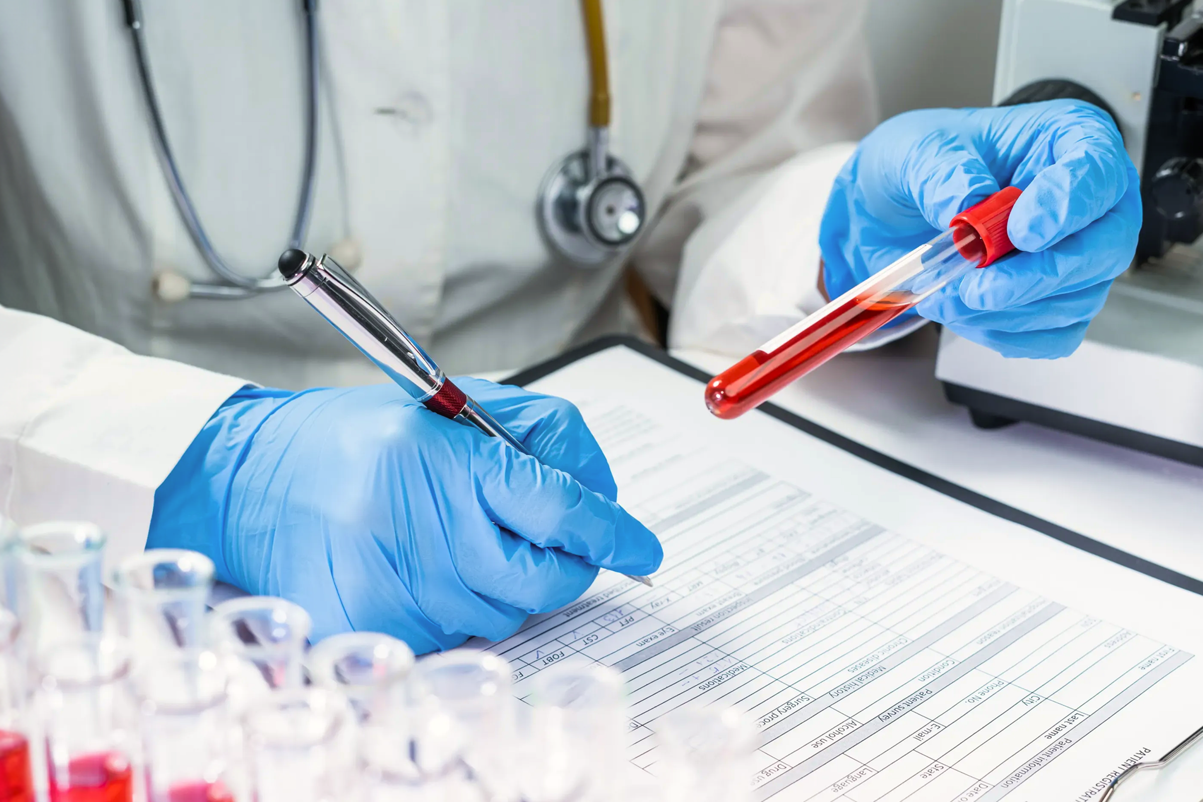
A study published in Nature Metabolism has generated significant attention with a finding that seems to upend conventional thinking about NAD+ and aging: whole blood NAD+ levels do not decline with age.¹ Some interpretations of this work have gone further, suggesting that age-related NAD+ decline may be a “myth.” Before accepting that conclusion, it is worth understanding what the study actually measured—and what it didn’t.
This article aims to explain what the study found, where its conclusions appropriately apply, and why the existing evidence for NAD+ decline with aging remains scientifically sound, and how this study is a refinement of the NAD+ aging hypothesis, not a refutation of it.
Understanding What Was—and Wasn’t—Measured
The researchers, Trętowicz and colleagues, used state-of-the-art analytical methods to measure NAD+ in the whole blood of participants across seven independent cohorts.1 They found that blood NAD+ remained remarkably stable across age groups and was largely unaffected by lifestyle interventions, though it did increase in response to nicotinamide riboside (NR) supplementation.
This is a meaningful contribution to the field. It suggests that whole blood NAD+ should not be used as a stand-alone biomarker for chronological aging, and that caution is warranted when interpreting blood-based measurements in clinical or consumer contexts.
However, the critical distinction that is easily lost in most coverage is: the study measured NAD+ in whole blood—not in the organs and tissues where age-related NAD+ biology may be most relevant. Whole blood is a complex matrix dominated by red blood cells and circulating immune cells. It may tell us relatively little about NAD+ status in the brain, skeletal muscle, skin, liver, or kidney—the compartments where the scientific case for age-related NAD+ decline is mostly built, and a distinction that was a central point of discussion at the recent NAD for Health conference in Copenhagen.
Concluding from stable whole blood NAD+ that NAD+ does not decline with aging anywhere in the body is the scientific equivalent of measuring the temperature outside your front door and concluding it must be the same temperature in every room of your house.
Three Methodological Considerations Worth Understanding
Beyond the tissue question, three specific methodological issues deserve attention.
- Lack of Red Blood Cell Normalization: The study did not adjust NAD+ measurements for red blood cell (RBC) count, hematocrit, or hemoglobin—values that normally decrease with age and tend to be lower in women than in men.² The authors did conduct exploratory analyses examining whether hematocrit confounded their NAD+ measurements, finding no significant association in two smaller sub-cohorts. However, they acknowledged these analyses “were not designed to formally test this relationship” and recommended future studies record hematocrit at the time of blood collection. Given the small samples involved, this question cannot be considered settled. This matters because NAD+ is present in all blood cells, and since RBCs make up roughly 40-45% of total blood volume and are by far the most abundant cell type, they represent a substantial portion of whole-blood NAD+.³ If RBC count falls with age but total blood NAD+ stays constant, the implication is that NAD+ per red blood cell is actually increasing with age, which is biologically implausible. For this reason, many research groups normalize RBC count precisely to avoid this interpretive problem, and the same logic applies when comparing results between men and women.
- Insufficient Sample Size: The study pooled several smaller observational studies and clinical trials rather than drawing from a single large cohort—an approach that introduces heterogeneity and limits the ability to detect modest but real biological effects. An absence of a detected effect is not the same as an absence of an effect. Dr. Howard Sesso, Associate Professor of Medicine at Harvard Medical School, put it directly:
“Their finding that NAD+ levels do not vary with age is provocative, but it is very difficult to make a definitive conclusion when combining several smaller underpowered observational studies and clinical trials. Any differences in NAD+ levels by age may simply be more modest and require larger numbers of participants in a single observational study or clinical trial.”
He also emphasized the broader need this study underscores:
“This study highlights how important it is for the NAD+ research community to understand not only how to optimally measure NAD+ levels in whole blood and/or tissues, but also the urgent need for gold-standard, long-term, large-scale randomized clinical trials testing interventions that impact NAD+ levels and subsequent healthy aging outcomes.”
- Pre-Analytical Variability Across Cohorts: The study combined seven cohorts collected under different conditions, introducing potential variability from factors such as collection tubes, processing delays, storage temperature, and freeze-thaw handling—all of which can substantially influence NAD+ measurements.⁴ The variation between cohorts in this study was approximately 10-15 nmol/mL—while the age-related effect being examined was only around 1.5 nmol/mL. When technical variation is nearly ten times the size of the biological signal, it is very difficult to draw confident conclusions in either direction.
A 2024 study by Pospieszna et al. illustrates the importance of controlling these variables.⁵ The researchers measured RBC NAD+ in 206 individuals aged 20-90, including elite endurance athletes, elite sprinters, and untrained controls, all processed under standardized conditions. RBC NAD+ declined significantly with age in every group. Although athletes consistently exhibited higher NAD+ levels than untrained individuals—supporting a link between physical activity and NAD+ status—the age-related decline remained evident even in the most highly trained cohort.
What the Human Tissue Evidence Actually Shows
The scientific case for age-related NAD+ decline does not rest on blood measurements. It rests primarily on direct tissue measurements and non-invasive imaging studies across multiple human tissues.
- Skin: A study by Massudi et al., 2012 measured NAD+ in non-sun-exposed pelvic skin from individuals ranging from newborn to 77 years of age.⁶ NAD+ was negatively correlated with age in both males and females—a straightforward finding from a tissue that is accessible and does not require major surgical intervention.
- Brain: Multiple studies using phosphorus and proton magnetic resonance spectroscopy (MRS)—a non-invasive imaging technique that can quantify NAD+ inside the living brain without biopsy—have documented age-related declines. Zhu et al., 2015 demonstrated decreases in NAD+ and the NAD+/NADH ratio in the occipital brain with aging.⁷ Bagga et al., 2020 replicated this finding using 7-Tesla MRS. These are direct, in vivo human tissue measurements.⁸
- Skeletal Muscle: Janssens et al., 2022 found that muscle NAD+ was lower in older adults than younger adults, and that physically trained older adults had levels closer to those of younger people.⁹ This is consistent with the protective role of physical activity seen across multiple studies, including the Pospieszna RBC data.
- Liver: Zhou et al., 2016 found that NAD+ levels in non-diseased liver tissue from patients over 60 were approximately 70% of the levels observed in patients under 45, consistent with an age-related decline in tissue NAD+.¹⁰
Tissues differ from blood in cell composition, NAD+ turnover rates, biosynthetic enzyme expression, and exposure to NAD+-consuming enzymes. Stable blood levels simply do not speak to what is happening in these compartments.
The Blood Picture Itself Is More Complicated Than One Study Can Resolve
It is also worth noting that the Trętowicz study is not the only study of its kind in this space. A study by Chaleckis et al. published in 2016 compared blood metabolites between young and elderly adults under well-controlled analytical conditions and found that NAD+ was significantly lower in the elderly group.¹¹ Similarly, in a 2023 study by Wang et al., the researchers found that whole blood NAD+ levels varied substantially across individuals, with clear differences tied to both age and sex.¹² Together, these studies make an important point: the blood signal is real, but heterogeneous—and detecting it reliably depends heavily on methodology, normalization, and cohort design.
Yang et al., 2022 added further nuance in a larger community-based cohort of 1,518 adults. Rather than a simple linear decline across the full population, men had significantly higher blood NAD+ than women overall, and men in the oldest age group showed a statistically significant age-associated decrease that women did not.¹³ Pooled, unstratified analyses can easily obscure these sex-specific and age-band-specific differences—and the absence of a detected effect is not the same as the absence of an effect.
What this body of evidence collectively suggests is not that blood NAD+ is meaningless, but that it is a more complicated signal than a single cross-cohort analysis can resolve. What the field needs is larger, longitudinal, sex-stratified research with standardized methodology—not a categorical conclusion in either direction.
What This Means for NAD+ Precursor Supplementation
The Trętowicz study itself confirms that whole blood NAD+ rises in response to NR supplementation—consistent with the broader clinical literature.¹⁴'¹⁵ This matters because the rationale for NAD+ precursor supplementation was never premised solely on blood measurements declining with age. It is built on tissue-level NAD+ biology and the demonstrated ability of precursors to augment NAD+ in relevant compartments. Emerging human data support this, including studies showing increases in brain and muscle NAD+ following supplementation.¹⁶⁻¹⁸
A stable whole blood signal with age does not change that framework.
Reframing the Question: Where Does NAD+ Decline with Age—and Why It Matters
The Trętowicz study is a useful contribution that should sharpen how the field thinks about blood-based NAD+ measurement. However, its findings are contradicted by several other studies; the Trętowicz study is not a refutation of tissue biology, and characterizing age-related NAD+ decline as a “myth” on this basis is not supported by the scientific evidence.
Given the totality of the evidence, a more accurate summary of where the science stands is as follows:
- Whole blood NAD+ may not be a reliable stand-alone biomarker for chronological aging.
- Direct human tissue measurements in skin, brain, skeletal muscle, and liver show consistent evidence of age-related NAD+ decline.
- Blood NAD+ associations with age are complex, likely sex-dependent, and cannot be resolved by any single study.
- NAD+ precursor supplementation raises blood NAD+ and shows emerging evidence of tissue-level effects in brain and muscle.
The more productive question—and the one the field is actively working to answer—is not whether NAD+ declines with age at all, but precisely where that decline occurs, in whom, over what timescale, and what its functional consequences are. That is a considerably more interesting scientific problem than any single headline can resolve.
References
- Trętowicz, M. M., Scantlebery, A. M. L., Schomakers, B. V., Eroğlu, K. D., Weeghel, M. van, Spek, V., Vinten, K. T., Legon, L., Coskun, E., Millan-Domingo, F., Olaso-Gonzalez, G., Gomez-Cabrera, M. C., Montoro-García, S., Noguera-Navarro, C., Kuilenburg, A. B. P. van, Moco, S., Hattum, J. C. van, Jørstad, H. T., Benali, M., … Houtkooper, R. H. (2026). Human whole-blood NAD+ levels do not vary with age or lifestyle interventions. Nature Metabolism, 1–9. https://doi.org/10.1038/s42255-026-01537-5
- Kyriacou, R. P., & Shibeeb, S. (2025). Red Blood Cells and Human Aging: Exploring Their Biomarker Potential. Diagnostics, 15(16), 1993. https://doi.org/10.3390/diagnostics15161993
- Chaleckis, R., Ebe, M., Pluskal, T., Murakami, I., Kondoh, H., & Yanagida, M. (2014). Unexpected similarities between the Schizosaccharomyces and human blood metabolomes, and novel human metabolites. Molecular BioSystems, 10(10), 2538–2551. https://doi.org/10.1039/c4mb00346b
- Bi, H., Guo, Z., Jia, X., Liu, H., Ma, L., & Xue, L. (2020). The key points in the pre-analytical procedures of blood and urine samples in metabolomics studies. Metabolomics, 16(6), 68. https://doi.org/10.1007/s11306-020-01666-2
- Pospieszna, B., Kusy, K., Slominska, E. M., Zieliński, J., & Ciekot-Sołtysiak, M. (2024). Erythrocyte nicotinamide adenine dinucleotide concentration is enhanced by systematic sports participation. BMC Sports Science, Medicine and Rehabilitation, 16(1), 216. https://doi.org/10.1186/s13102-024-00999-y
- Massudi, H., Grant, R., Braidy, N., Guest, J., Farnsworth, B., & Guillemin, G. J. (2012). Age-Associated Changes In Oxidative Stress and NAD+ Metabolism In Human Tissue. PLoS ONE, 7(7), e42357. https://doi.org/10.1371/journal.pone.0042357
- Zhu, X.-H., Lu, M., Lee, B.-Y., Ugurbil, K., & Chen, W. (2015). In vivo NAD assay reveals the intracellular NAD contents and redox state in healthy human brain and their age dependences. Proceedings of the National Academy of Sciences, 112(9), 2876–2881. https://doi.org/10.1073/pnas.1417921112
- Bagga, P., Hariharan, H., Wilson, N. E., Beer, J. C., Shinohara, R. T., Elliott, M. A., Baur, J. A., Marincola, F. M., Witschey, W. R., Haris, M., Detre, J. A., & Reddy, R. (2020). Single‐Voxel 1H MR spectroscopy of cerebral nicotinamide adenine dinucleotide (NAD+) in humans at 7T using a 32‐channel volume coil. Magnetic Resonance in Medicine, 83(3), 806–814. https://doi.org/10.1002/mrm.27971
- Janssens, G. E., Grevendonk, L., Perez, R. Z., Schomakers, B. V., Bosch, J. de V. den, Geurts, J. M. W., Weeghel, M. van, Schrauwen, P., Houtkooper, R. H., & Hoeks, J. (2022). Healthy aging and muscle function are positively associated with NAD+ abundance in humans. Nature Aging, 2(3), 254–263. https://doi.org/10.1038/s43587-022-00174-3
- Zhou, C., Yang, X., Hua, X., Liu, J., Fan, M., Li, G., Song, J., Xu, T., Li, Z., Guan, Y., Wang, P., & Miao, C. (2016). Hepatic NAD+ deficiency as a therapeutic target for non‐alcoholic fatty liver disease in ageing. British Journal of Pharmacology, 173(15), 2352–2368. https://doi.org/10.1111/bph.13513
- Chaleckis, R., Murakami, I., Takada, J., Kondoh, H., & Yanagida, M. (2016). Individual variability in human blood metabolites identifies age-related differences. Proceedings of the National Academy of Sciences, 113(16), 4252–4259. https://doi.org/10.1073/pnas.1603023113
- Wang, P., Chen, M., Hou, Y., Luan, J., Liu, R., Chen, L., Hu, M., & Yu, Q. (2023). Fingerstick blood assay maps real‐world NAD+ disparity across gender and age. Aging Cell. https://doi.org/10.1111/acel.13965
- Yang, F., Deng, X., Yu, Y., Luo, L., Chen, X., Zheng, J., Qiu, Y., Xiao, F., Xie, X., Zhao, Y., Guo, J., Hu, F., Zhang, X., Ju, Z., & Zhou, Y. (2022). Association of Human Whole Blood NAD+ Contents With Aging. Frontiers in Endocrinology, 13, 829658. https://doi.org/10.3389/fendo.2022.829658
- Conze, D., Brenner, C., & Kruger, C. L. (2019). Safety and Metabolism of Long-term Administration of NIAGEN (Nicotinamide Riboside Chloride) in a Randomized, Double-Blind, Placebo-controlled Clinical Trial of Healthy Overweight Adults. Scientific Reports, 9(1), 9772. https://doi.org/10.1038/s41598-019-46120-z
- Martens, C. R., Denman, B. A., Mazzo, M. R., Armstrong, M. L., Reisdorph, N., McQueen, M. B., Chonchol, M., & Seals, D. R. (2018). Chronic nicotinamide riboside supplementation is well-tolerated and elevates NAD+ in healthy middle-aged and older adults. Nature Communications, 9(1), 1286. https://doi.org/10.1038/s41467-018-03421-7
- Brakedal, B., Dölle, C., Riemer, F., Ma, Y., Nido, G. S., Skeie, G. O., Craven, A. R., Schwarzlmüller, T., Brekke, N., Diab, J., Sverkeli, L., Skjeie, V., Varhaug, K., Tysnes, O.-B., Peng, S., Haugarvoll, K., Ziegler, M., Grüner, R., Eidelberg, D., & Tzoulis, C. (2022). The NADPARK study: A randomized phase I trial of nicotinamide riboside supplementation in Parkinson’s disease. Cell Metabolism, 34(3), 396-407.e6. https://doi.org/10.1016/j.cmet.2022.02.001
- Nanga, R. P. R., Wiers, C. E., Elliott, M. A., Wilson, N. E., Liu, F., Cao, Q., Swago, S., Jacobs, P. S., Armbruster, R., Reddy, D., Baur, J. A., Witschey, W. R., Detre, J. A., & Reddy, R. (2024). Acute nicotinamide riboside supplementation increases human cerebral NAD+ levels in vivo. Magnetic Resonance in Medicine, 92(6), 2284–2293. https://doi.org/10.1002/mrm.30227
- Lapatto, H. A. K., Kuusela, M., Heikkinen, A., Muniandy, M., Kolk, B. W. van der, Gopalakrishnan, S., Pöllänen, N., Sandvik, M., Schmidt, M. S., Heinonen, S., Saari, S., Kuula, J., Hakkarainen, A., Tampio, J., Saarinen, T., Taskinen, M.-R., Lundbom, N., Groop, P.-H., Tiirola, M., … Pirinen, E. (2023). Nicotinamide riboside improves muscle mitochondrial biogenesis, satellite cell differentiation, and gut microbiota in a twin study. Science Advances, 9(2), eadd5163. https://doi.org/10.1126/sciadv.add5163