The Role of NAD+ in Immune Health and Function
Key Takeaways
- NAD+ is a foundational molecule for immune cells: It fuels cellular energy production, DNA repair, and stress-response pathways that help immune cells function in states of infection, inflammation, and oxidative damage.
- NAD+ supports both innate and adaptive immunity: It’s a key molecular foundation for healthy aging and balanced immune responses.
- Immune stress and viral infections deplete NAD+: This can impair immune function, cellular repair, and inflammatory regulation.
- NAD+ precursors can help: NR, NAM, and NMN can help restore NAD+, supporting immune cell resilience and function.
- Maintaining NAD+ works best alongside lifestyle factors: Balanced nutrition, exercise, sleep, and stress management all support immune health.
The immune system is a complex, highly coordinated network designed to protect the body from external threats—such as viruses, bacteria, and other pathogens—while also maintaining internal balance through constant surveillance and repair.¹ When immune function is compromised by aging, chronic stress, or infection, the consequences can extend well beyond acute illness, influencing recovery and long-term health outcomes.²
For decades, the public has closely associated immune health with specific nutrients—vitamin C for antioxidant defense and immune cell activity,³ zinc for immune cell development and communication,⁴ and vitamin D for the regulation of immune signaling and inflammatory responses.⁵ Other minerals also play important roles in immunity, with selenium supporting antioxidant enzyme function and antiviral defense,⁶ and copper contributing to immune cell maturation and oxidative stress regulation.⁷
In more recent years, other scientifically supported compounds have also gained attention for their roles in immune support. Probiotics, for example, can influence immune activity through interactions with the gut microbiome,⁸ while functional mushrooms contain bioactive polysaccharides that can modulate immune responses.⁹ Together, these advances reflect a growing understanding that immune health does not depend on a single nutrient or pathway, but rather on an interconnected network of cellular processes that support immune cell function and resilience.
Within this expanding scientific landscape, nicotinamide adenine dinucleotide (NAD+) has emerged as a critical but often underrecognized component of immune health. NAD+ is a vital molecule found in every living cell, where it supports cellular energy production, DNA repair, mitochondrial function, and the activity of NAD-dependent enzymes that coordinate cellular responses to metabolic, oxidative, and inflammatory stress.¹⁰'¹¹ NAD+ levels naturally decline with age¹² and can be further depleted by physiological stressors, including immune activation and viral infection. A growing body of research now suggests that NAD+ availability may influence both innate and adaptive immune responses, affecting how immune cells respond to stress, proliferate, and recover.
This article explores the evolving science linking NAD+ to immune health, including how innate and adaptive immunity differ, how immune stressors such as viral infections can impact NAD+ levels, and what current research suggests about the role of NAD+ and NAD-dependent pathways in immune cell function.
Understanding the Immune System: Innate vs. Adaptive Immunity
The immune system has two main and interconnected branches: innate immunity and adaptive immunity.¹³ While they differ in specificity, speed, and function, these systems work closely together to detect threats, limit damage, and establish long-lasting protection against future infections.
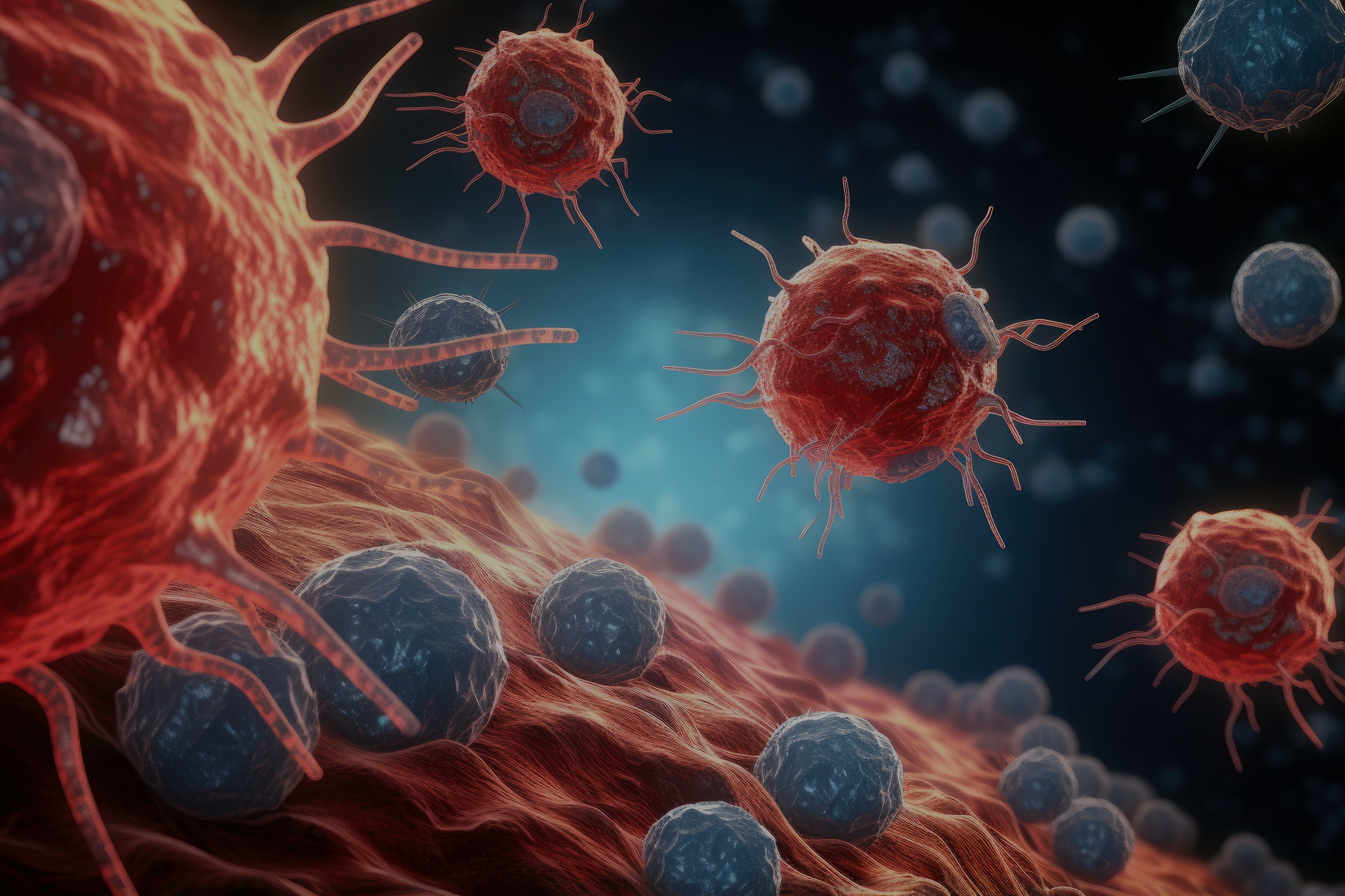
Innate immunity is the body’s first line of defense.¹⁴ It responds rapidly and non-specifically, targeting any perceived threat within minutes to hours of exposure. This branch includes physical barriers such as the skin and mucosal surfaces, as well as specialized immune cells—macrophages, neutrophils, dendritic cells, and natural killer (NK) cells—that detect and respond to signs of infection or cellular stress. Upon activation, these innate immune cells rapidly increase their metabolic activity to support functions such as pathogen engulfment (a process known as phagocytosis), inflammatory signaling, and antimicrobial molecule production.¹⁵ These immediate defense mechanisms place substantial demands on cellular energy production and stress response pathways.¹⁶
Conversely, adaptive immunity develops more slowly but provides highly specific and long-lasting protection.¹⁷ This branch of the immune system is mediated primarily by T cells and B cells, which recognize and remember distinct features of pathogens. Once activated, adaptive immune cells undergo extensive proliferation and differentiation, producing effector cells that eliminate infected targets and memory cells that enable faster and more efficient responses upon re-exposure.¹⁸
Both innate and adaptive immunity rely on cells that must rapidly adapt to changing conditions, placing exceptionally high energy and repair demands, from rapid inflammatory responses to long-term immune memory formation.¹⁹ Effective immunity depends on sustained energy and robust cellular function—a context in which NAD+ plays a central role.
What Is NAD+ and How Does It Support Immune Health?
NAD+ is a critical molecule found in all living cells—including all immune cells—where it helps regulate processes essential for maintaining health. At its core, NAD+ supports cellular energy production, DNA repair, mitochondrial function, and cell signaling, providing the foundation that enables immune cells—particularly macrophages, neutrophils, T cells, and B cells—to respond effectively to stress or infection.
Immune cells depend on NAD+ to meet these demands.²⁰ Without enough NAD+, they may struggle to generate the energy required for activation, repair DNA damage caused by oxidative stress, and regulate inflammatory responses. In other words, the availability of NAD+ can directly shape how effectively immune cells can perform their functions.
Two families of NAD-dependent enzymes are central to these processes: sirtuins and poly (ADP-ribose) polymerases (PARPs). PARPs use NAD+ to detect and repair DNA damage and to help control inflammation,²¹ while sirtuins regulate inflammatory signaling, manage cellular stress responses, and influence immune cell differentiation and health.²²
When NAD+ levels drop, the activity of both PARPs and sirtuins diminishes.²³ Immune cells may then mount weaker responses, repair damage less efficiently, and fail to control inflammation properly.²⁰ In this way, NAD+ serves as a central metabolic link between immunity, inflammation, and recovery—essentially acting as the metabolic wiring that enables energy production and cellular repair, allowing immune cells to function optimally.
Even modest decreases in NAD+ can have significant consequences. Reduced sirtuin activity can impair T cell function during periods of severe stress or infection,²⁴ while disruptions in mitochondrial function—which also depends on NAD+—can exhaust T cells and limit their ability to respond effectively to pathogens.²⁵ Together, these findings highlight the essential role of NAD+ in maintaining energy balance, supporting DNA repair, and enabling immune cells to mount coordinated and effective responses.
How Viral Infections and Immune Stress Deplete NAD+ Levels
When a virus infects a cell, it hijacks the host’s machinery to replicate, triggering a surge in the cell’s defense systems.²⁶ This creates immune stress, a heightened demand for energy and repair driven by infection, chronic inflammation, or other immune challenges. To counter the threat, cells activate anti-viral defense mechanisms that dramatically increase NAD+ consumption, placing significant strain on metabolic and repair processes.²⁷
Central to this response are NAD-dependent enzymes, particularly PARPs and sirtuins, which are mobilized during infection to counteract viral replication and repair cellular damage.²⁸ PARPs are rapidly upregulated to detect and repair DNA damage—a process that consumes large amounts of NAD+.²¹ Similarly, sirtuins increase their activity to regulate inflammatory signaling and stress responses, further depleting NAD+ stores.²⁷ When activation of these enzymes is prolonged or excessive, NAD+ levels can be rapidly drained, creating a metabolic bottleneck for immune cells.²⁷
Evidence from both human and preclinical studies demonstrates the scope of this depletion. In people with COVID-19, NAD+ metabolism is disrupted, and SIRT1 activity is altered, with changes more pronounced in people with severe disease.²⁹ Compared to healthy controls, COVID-19 patients also exhibit slightly lower NAD+ levels alongside increased NAD+ turnover, suggesting that infection drives both accelerated consumption and compensatory production of NAD+ in an effort to meet cellular demand.³⁰ In mouse models of COVID-19, NAD+ levels in primary immune cells were reduced by over threefold, primarily due to upregulated PARP activity, further supporting the notion that respiratory infection drives NAD+ consumption.³¹
This pattern extends beyond COVID-19. HSV-1 (herpes simplex virus-1) infection can cause a dramatic drop in cellular NAD+ levels—up to an 80% reduction in some cases—leaving infected cells with only a fraction of their normal NAD+ stores.³² HIV-1 infection similarly decreases intracellular NAD+, with severity varying by viral load and strain.³³ Even in chronic immune stress—such as in T cells from patients with psoriasis—the NAD+/NADH ratio is reduced, suggesting that prolonged immune activation alone is sufficient to deplete NAD+.³⁴
This depletion carries significant functional consequences. Without adequate NAD+, immune cells struggle to maintain the energy production required for robust immune responses and cannot repair DNA damage efficiently.²⁰
Together, these findings reveal a vicious cycle: immune stress and viral infection activate NAD-consuming enzymes—particularly PARPs—to repair damage and counteract viral replication, which rapidly depletes cellular NAD+ stores. As NAD+ levels decline, immune cells lose metabolic efficiency, DNA repair slows, and inflammatory responses can become dysregulated, further compromising immune defense and potentially prolonging illness. This cycle underscores why ensuring sufficient NAD+ availability is essential for sustaining immune cell function and supporting recovery during viral stress.
Increasing NAD+ Levels May Support Immune Health
While NAD+ is not an immune stimulant—it does not directly activate immune cells—it is a foundational support molecule that provides the metabolic infrastructure for immune cells to function at their best. By maintaining adequate NAD+ levels, cells can sustain energy production, repair DNA efficiently, and regulate inflammatory signaling, which are all essential for healthy immune functioning.
There are several ways to support NAD+ levels, including lifestyle approaches like moderate exercise and caloric restriction or intermittent fasting, which enhance NAD+ availability by promoting metabolic efficiency. Interventions that increase NAD+ availability—such as precursors like nicotinamide riboside (NR), nicotinamide (NAM), or nicotinamide mononucleotide (NMN)—can help cells meet the intense demands of viral infection and immune stress.
NAD+ Support During Infection and Pathogen Defense
Preclinical research demonstrates how restoring NAD+ availability can help break the cycle of infection-driven depletion. Preclinical research demonstrates how increasing NAD+ availability can help immune cells maintain their defensive capabilities during infection. In mouse models of coronavirus infection, NR supplementation provided immune cells with the metabolic support needed to limit viral replication: in mouse fibroblasts infected with murine hepatitis virus (a coronavirus closely related to SARS-CoV-2), NR reduced viral replication more than sixfold, and in macrophages, replication decreased nearly threefold.³¹ In a separate study looking at viral recovery, mice treated with NR maintained higher body weight during recovery, reflecting not just cellular resilience but improved overall systemic health.³⁵
This antiviral support appears to work through multiple mechanisms. Research shows that NR activates the Citric Acid cycle (also known as the Krebs cycle)—the cell’s central metabolic engine—improving energy production capacity while simultaneously limiting the resources available for viral replication.³⁶ Similar patterns emerge with bacterial infections. In models of bacterial pneumonia, restoring NAD+ through supplementation with NR, NMN, or NAM increased intracellular NAD+ levels in bronchial epithelial cells and directly reduced bacterial growth, demonstrating a direct antibacterial effect.³⁷
Perhaps most striking are findings from sepsis models—severe, life-threatening systemic infections where the immune system faces overwhelming challenges. NR supplementation restored NAD+ and sirtuin activity in T cells, reduced bacterial load in lungs and bloodstream, protected organs from inflammatory damage, and significantly decreased mortality rates.³⁸ These studies illustrate a consistent principle: by maintaining the metabolic foundation that immune cells depend on, NAD+ precursors help sustain cellular defenses during the intense demands of infection.
Enhancing Immune Cell Function and Reducing Inflammation
Beyond acute infection response, NAD+ plays a crucial role in maintaining the long-term health and responsiveness of immune cells, particularly when those cells face chronic stress or dysfunction. This is especially evident in T cells, the adaptive immune cells responsible for targeting infected or malignant cells.
Tumor-infiltrating T cells offer a clear example of how NAD+ support can restore immune cell function. These immune cells often become functionally impaired in the tumor environment, accumulating damaged mitochondria and entering a state of terminal exhaustion where they can no longer mount effective responses.³⁹
NR supplementation improves mitochondrial health in these exhausted T cells, restores their responsiveness, and enhances their cytotoxic activity against cancer cells.³⁹ Similarly, NR treatment has been shown to inhibit T cell exhaustion, increase their ability to kill infected or malignant cells, and reduce immune cell death—all by supporting mitochondrial health. During sepsis, NR has not only restored NAD+ levels but has also prevented the severe depletion of T cells and other white blood cells in the spleen, helping maintain a functional immune cell population.³⁸
NAD+ support also appears to modulate inflammatory signaling in ways that may benefit people with overactive immune responses. In monocytes from individuals with systemic lupus erythematosus—an autoimmune condition characterized by chronic inflammation—NR reduced expression of inflammatory cytokines and lessened type-I interferon signaling, key mediators of inflammation, while increasing NAD+ metabolite levels in the bloodstream.⁴⁰ Similarly, NR improved the resilience of CD4+ T cells from patients with psoriasis, enhancing their resistance to oxidative stress and lowering hyperactive immune responses.³⁴ These findings suggest that by supporting cellular metabolism and energy balance, NAD+ precursors may help immune cells respond more appropriately, avoiding both underactivity and excessive inflammation.
Preclinical studies with other NAD+ precursors, such as NAM and NMN,³⁷'⁴¹ show comparable effects, highlighting that multiple pathways to increasing NAD+ availability—whether through NR, NMN, or NAM—may contribute to immune cell resilience and balanced function.
Together, these findings highlight a consistent pattern: NAD+ acts as the metabolic backbone of immune cells: it helps fuel cellular energy needs, supports DNA repair and mitochondrial function, regulates inflammation, and enhances resilience to both pathogens and chronic stress. That said, this support is most effective as part of a broader foundation of health that includes adequate sleep, stress management, balanced nutrition, and other essential practices. While preclinical evidence and emerging clinical evidence are promising, additional human studies are still needed to fully understand optimal dosing and considerations for supplementing with NAD+ precursors in the context of immune health.
NAD+ as a Foundation for Immune Health and Longevity
NAD+ plays a multifaceted role in immune health, supporting cellular energy production, DNA repair, and the regulation of inflammation. Through these actions, NAD+ provides the molecular infrastructure that enables immune cells to respond effectively to threats and pathogens. Both branches of the immune system—innate immunity, which mounts immediate and physical defenses, and adaptive immunity, which provides specific, long-lasting protection—depend on NAD+ to meet their high metabolic and repair demands.
Immune stress from infection, chronic inflammation, or other challenges can rapidly deplete NAD+, underscoring its critical role in sustaining healthy immune function. Key NAD-dependent enzymes, like PARPs and sirtuins, orchestrate DNA repair, stress responses, and antiviral defense—but their activity consumes NAD+, creating a delicate balance between immune activation and cellular resources.
NAD+ does not act in isolation. Its effects are intertwined with other aspects of a healthy lifestyle, including nutrition, exercise, sleep, and stress management. Vitamins, minerals, and metabolites such as vitamin C, zinc, vitamin D, probiotics, and certain functional mushrooms can all contribute to immune resilience, working alongside NAD+ to support energy metabolism, repair processes, and regulated immune signaling.
Supporting NAD+ levels through these healthy lifestyle practices—or in combination with supplementation with NAD+ precursors like NR, NAM, or NMN—may help maintain immune cell function, support recovery, and enhance resilience during periods of stress or infection. While NAD+ is not a magic solution, it serves as a central foundation for healthy aging and balanced immunity, providing an essential metabolic backbone that allows the immune system to function optimally.
References
- Medina, K. L. (2016). Chapter 4 Overview of the immune system. Handbook of Clinical Neurology, 133, 61–76. https://doi.org/10.1016/b978-0-444-63432-0.00004-9
- Goyani, P., Christodoulou, R., & Vassiliou, E. (2024). Immunosenescence: Aging and Immune System Decline. Vaccines, 12(12), 1314. https://doi.org/10.3390/vaccines12121314
- Carr, A. C., & Maggini, S. (2017). Vitamin C and Immune Function. Nutrients, 9(11), 1211. https://doi.org/10.3390/nu9111211
- Wessels, I., Fischer, H. J., & Rink, L. (2021). Dietary and Physiological Effects of Zinc on the Immune System. Annual Review of Nutrition, 41(1), 1–43. https://doi.org/10.1146/annurev-nutr-122019-120635
- Sîrbe, C., Rednic, S., Grama, A., & Pop, T. L. (2022). An Update on the Effects of Vitamin D on the Immune System and Autoimmune Diseases. International Journal of Molecular Sciences, 23(17), 9784. https://doi.org/10.3390/ijms23179784
- Tinggi, U. (2008). Selenium: its role as antioxidant in human health. Environmental Health and Preventive Medicine, 13(2), 102–108. https://doi.org/10.1007/s12199-007-0019-4
- Percival, S. (1998). Copper and immunity. The American Journal of Clinical Nutrition, 67(5), 1064S-1068S. https://doi.org/10.1093/ajcn/67.5.1064s
- Mazziotta, C., Tognon, M., Martini, F., Torreggiani, E., & Rotondo, J. C. (2023). Probiotics Mechanism of Action on Immune Cells and Beneficial Effects on Human Health. Cells, 12(1), 184. https://doi.org/10.3390/cells12010184
- Zhao, S., Gao, Q., Rong, C., Wang, S., Zhao, Z., Liu, Y., & Xu, J. (2020). Immunomodulatory Effects of Edible and Medicinal Mushrooms and Their Bioactive Immunoregulatory Products. Journal of Fungi, 6(4), 269. https://doi.org/10.3390/jof6040269
- Covarrubias, A. J., Perrone, R., Grozio, A., & Verdin, E. (2021). NAD+ metabolism and its roles in cellular processes during ageing. Nature Reviews Molecular Cell Biology, 22(2), 119–141. https://doi.org/10.1038/s41580-020-00313-x
- Campagna, R., & Vignini, A. (2023). NAD+ Homeostasis and NAD+-Consuming Enzymes: Implications for Vascular Health. Antioxidants, 12(2), 376. https://doi.org/10.3390/antiox12020376
- Massudi, H., Grant, R., Braidy, N., Guest, J., Farnsworth, B., & Guillemin, G. J. (2012). Age-Associated Changes In Oxidative Stress and NAD+ Metabolism In Human Tissue. PLoS ONE, 7(7), e42357. https://doi.org/10.1371/journal.pone.0042357
- Wang, R., Lan, C., Benlagha, K., Camara, N. O. S., Miller, H., Kubo, M., Heegaard, S., Lee, P., Yang, L., Forsman, H., Li, X., Zhai, Z., & Liu, C. (2024). The interaction of innate immune and adaptive immune system. MedComm, 5(10), e714. https://doi.org/10.1002/mco2.714
- Hillion, S., Arleevskaya, M. I., Blanco, P., Bordron, A., Brooks, W. H., Cesbron, J. Y., Kaveri, S., Vivier, E., & Renaudineau, Y. (2020). The Innate Part of the Adaptive Immune System. Clinical Reviews in Allergy & Immunology, 58(2), 151–154. https://doi.org/10.1007/s12016-019-08740-1
- Takashi, A., Junkichi, Y., Shinichi, O., Mitsuhisa, F., Masataka, K., Hiroaki, K., Shin, I., & Katuhisa, I. (n.d.). Antegrade Bipedicled Submental Island Flap for Anterior Oropharyngeal Defect Reconstruction after Ablative Surgery. Otology & Rhinology. https://doi.org/10.4172/2324-8785.s1-016
- Delmastro-Greenwood, M. M., & Piganelli, J. D. (2013). Changing the energy of an immune response. American Journal of Clinical and Experimental Immunology, 2(1), 30–54.
- Bonilla, F. A., & Oettgen, H. C. (2010). Adaptive immunity. Journal of Allergy and Clinical Immunology, 125(2), S33–S40. https://doi.org/10.1016/j.jaci.2009.09.017
- Mandujano-Tinoco, E. A., Sultan, E., Ottolenghi, A., Gershoni-Yahalom, O., & Rosental, B. (2021). Evolution of Cellular Immunity Effector Cells; Perspective on Cytotoxic and Phagocytic Cellular Lineages. Cells, 10(8), 1853. https://doi.org/10.3390/cells10081853
- Faas, M. M., & Vos, P. de. (2020). Mitochondrial function in immune cells in health and disease. Biochimica et Biophysica Acta (BBA) - Molecular Basis of Disease, 1866(10), 165845. https://doi.org/10.1016/j.bbadis.2020.165845
- Mann, R., Stavrou, V., & Dimeloe, S. (2025). NAD + metabolism and function in innate and adaptive immune cells. Journal of Inflammation, 22(1), 30. https://doi.org/10.1186/s12950-025-00457-7
- Murata, M. M., Kong, X., Moncada, E., Chen, Y., Imamura, H., Wang, P., Berns, M. W., Yokomori, K., & Digman, M. A. (2019). NAD+ consumption by PARP1 in response to DNA damage triggers metabolic shift critical for damaged cell survival. Molecular Biology of the Cell, 30(20), 2584–2597. https://doi.org/10.1091/mbc.e18-10-0650
- Hamaidi, I., & Kim, S. (2022). Sirtuins are crucial regulators of T cell metabolism and functions. Experimental & Molecular Medicine, 54(3), 207–215. https://doi.org/10.1038/s12276-022-00739-7
- Imai, S., & Guarente, L. (2014). NAD+ and sirtuins in aging and disease. Trends in Cell Biology, 24(8), 464–471. https://doi.org/10.1016/j.tcb.2014.04.002
- Zhao, G., Yang, X., Zhang, C., Dong, W., Dong, F., Zhang, J., Chen, X.-Y., Yao, R., Xiao, Z., Chen, L., Yao, Y., & Lu, Z. (2023). SUPPLEMENTATION WITH NICOTINAMIDE RIBOSIDE ATTENUATES T CELL EXHAUSTION AND IMPROVES SURVIVAL IN SEPSIS. Shock, 60(2), 238–247. https://doi.org/10.1097/shk.0000000000002153
- Yu, Y.-R., Imrichova, H., Wang, H., Chao, T., Xiao, Z., Gao, M., Rincon-Restrepo, M., Franco, F., Genolet, R., Cheng, W.-C., Jandus, C., Coukos, G., Jiang, Y.-F., Locasale, J. W., Zippelius, A., Liu, P.-S., Tang, L., Bock, C., Vannini, N., & Ho, P.-C. (2020). Disturbed mitochondrial dynamics in CD8+ TILs reinforce T cell exhaustion. Nature Immunology, 21(12), 1540–1551. https://doi.org/10.1038/s41590-020-0793-3
- Wu, W., Luo, X., & Ren, M. (2022). Clearance or Hijack: Universal Interplay Mechanisms Between Viruses and Host Autophagy From Plants to Animals. Frontiers in Cellular and Infection Microbiology, 11, 786348. https://doi.org/10.3389/fcimb.2021.786348
- Shang, J., Smith, M. R., Anmangandla, A., & Lin, H. (2021). NAD+-consuming enzymes in immune defense against viral infection. Biochemical Journal, 478(23), 4071–4092. https://doi.org/10.1042/bcj20210181
- Groth, B., Venkatakrishnan, P., & Lin, S.-J. (2021). NAD+ Metabolism, Metabolic Stress, and Infection. Frontiers in Molecular Biosciences, 8, 686412. https://doi.org/10.3389/fmolb.2021.686412
- Lonati, C., Berezhnoy, G., Lawler, N., Masuda, R., Kulkarni, A., Sala, S., Nitschke, P., Zizmare, L., Bucci, D., Cannet, C., Schäfer, H., Singh, Y., Gray, N., Lodge, S., Nicholson, J., Merle, U., Wist, J., & Trautwein, C. (2024). Urinary phenotyping of SARS-CoV-2 infection connects clinical diagnostics with metabolomics and uncovers impaired NAD+ pathway and SIRT1 activation. Clinical Chemistry and Laboratory Medicine (CCLM), 62(4), 770–788. https://doi.org/10.1515/cclm-2023-1017
- Valderrábano, R. J., Wipper, B., Pencina, K. M., Migaud, M., Shang, Y. V., Latham, N. K., Montano, M., Cunningham, J. M., Wilson, L., Peng, L., Memish‐Beleva, Y., Bhargava, A., Swain, P. M., Lehman, P., Lavu, S., Livingston, D. J., & Bhasin, S. (2024). Dysregulated nicotinamide adenine dinucleotide metabolome in patients hospitalized with COVID‐19. Aging Cell. https://doi.org/10.1111/acel.14326
- Heer, C. D., Sanderson, D. J., Voth, L. S., Alhammad, Y. M. O., Schmidt, M. S., Trammell, S. A. J., Perlman, S., Cohen, M. S., Fehr, A. R., & Brenner, C. (2020). Coronavirus infection and PARP expression dysregulate the NAD metabolome: An actionable component of innate immunity. Journal of Biological Chemistry, 295(52), 17986–17996. https://doi.org/10.1074/jbc.ra120.015138
- Grady, S. L., Hwang, J., Vastag, L., Rabinowitz, J. D., & Shenk, T. (2012). Herpes Simplex Virus 1 Infection Activates Poly(ADP-Ribose) Polymerase and Triggers the Degradation of Poly(ADP-Ribose) Glycohydrolase. Journal of Virology, 86(15), 8259–8268. https://doi.org/10.1128/jvi.00495-12
- Murray, M. F., Nghiem, M., & Srinivasan, A. (1995). HIV Infection Decreases Intracellular Nicotinamide Adenine Dinucleotide [NAD]. Biochemical and Biophysical Research Communications, 212(1), 126–131. https://doi.org/10.1006/bbrc.1995.1945
- Han, K., Singh, K., Meadows, A. M., Sharma, R., Hassanzadeh, S., Wu, J., Goss-Holmes, H., Huffstutler, R. D., Teague, H. L., Mehta, N. N., Griffin, J. L., Tian, R., Traba, J., & Sack, M. N. (2023). Boosting NAD preferentially blunts Th17 inflammation via arginine biosynthesis and redox control in healthy and psoriasis subjects. Cell Reports Medicine, 4(9), 101157. https://doi.org/10.1016/j.xcrm.2023.101157
- Izadpanah, A., Mudd, J. C., Garcia, J. G. N., Srivastav, S., Abdel-Mohsen, M., Palmer, C., Goldman, A. R., Kolls, J. K., Qin, X., & Rappaport, J. (2023). SARS-CoV-2 infection dysregulates NAD metabolism. Frontiers in Immunology, 14, 1158455. https://doi.org/10.3389/fimmu.2023.1158455
- Lee, S. R., Roh, J. Y., Ryu, J., Shin, H.-J., & Hong, E.-J. (2022). Activation of TCA cycle restrains virus-metabolic hijacking and viral replication in mouse hepatitis virus-infected cells. Cell & Bioscience, 12(1), 7. https://doi.org/10.1186/s13578-021-00740-z
- Klabunde, B., Wesener, A., Bertrams, W., Beinborn, I., Paczia, N., Surmann, K., Blankenburg, S., Wilhelm, J., Serrania, J., Knoops, K., Elsayed, E. M., Laakmann, K., Jung, A. L., Kirschbaum, A., Hammerschmidt, S., Alshaar, B., Gisch, N., Mraheil, M. A., Becker, A., … Schmeck, B. (2023). NAD+ metabolism is a key modulator of bacterial respiratory epithelial infections. Nature Communications, 14(1), 5818. https://doi.org/10.1038/s41467-023-41372-w
- Zhao, G., Yang, X., Zhang, C., Dong, W., Dong, F., Zhang, J., Chen, X.-Y., Yao, R., Xiao, Z., Chen, L., Yao, Y., & Lu, Z. (2023). SUPPLEMENTATION WITH NICOTINAMIDE RIBOSIDE ATTENUATES T CELL EXHAUSTION AND IMPROVES SURVIVAL IN SEPSIS. Shock, 60(2), 238–247. https://doi.org/10.1097/shk.0000000000002153
- Yu, Y.-R., Imrichova, H., Wang, H., Chao, T., Xiao, Z., Gao, M., Rincon-Restrepo, M., Franco, F., Genolet, R., Cheng, W.-C., Jandus, C., Coukos, G., Jiang, Y.-F., Locasale, J. W., Zippelius, A., Liu, P.-S., Tang, L., Bock, C., Vannini, N., & Ho, P.-C. (2020). Disturbed mitochondrial dynamics in CD8+ TILs reinforce T cell exhaustion. Nature Immunology, 21(12), 1540–1551. https://doi.org/10.1038/s41590-020-0793-3
- Wu, J., Singh, K., Lin, A., Meadows, A. M., Wu, K., Shing, V., Bley, M., Hassanzadeh, S., Huffstutler, R. D., Schmidt, M. S., Blanco, L. P., Tian, R., Brenner, C., Pirooznia, M., Kaplan, M. J., & Sack, M. N. (2022). Boosting NAD+ blunts toll-like receptor-4 induced type-I interferon in control and systemic lupus erythematosus monocytes. Journal of Clinical Investigation, 132(5), e139828. https://doi.org/10.1172/jci139828
- Kim, H.-W., Ryoo, G.-H., Jang, H.-Y., Rah, S.-Y., Lee, D. H., Kim, D.-K., Bae, E. J., & Park, B.-H. (2022). NAD+-boosting molecules suppress mast cell degranulation and anaphylactic responses in mice. Theranostics, 12(7), 3316–3328. https://doi.org/10.7150/thno.69684